More Pain Management & Rehabilitation Articles
Mild Degenerative Changes of the SI Joints: What the Report Means
By Your Health Magazine Health Information Team
Mild degenerative changes of the SI joints means an imaging test found small signs of wear in the sacroiliac joints, where the lower spine connects to the pelvis. This is usually not an emergency, does not necessarily cause pain, and does not by itself establish a diagnosis. Your symptoms, physical examination, age, medical history, and the rest of the imaging report determine whether the finding is clinically important.
What Are the SI Joints?
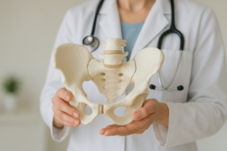
You have two sacroiliac, or SI, joints. They connect the sacrum at the bottom of the spine to the ilium bones on each side of the pelvis. These strong, weight-bearing joints allow only a small amount of movement while transferring force between the upper body and legs.
Like other joints, SI joints can change over time. Cartilage may thin, bone may become denser near the joint, and small bone spurs may develop. These findings fall within the broader category of joint disorders described by MedlinePlus.
What “Mild Degenerative Changes” May Include
“Mild” generally indicates limited or early findings rather than severe joint damage. The exact meaning can vary with the imaging method and the radiologist’s interpretation. A report may mention one or more of the following:
| Report term | What it generally means |
|---|---|
| Subchondral sclerosis | Increased bone density beneath the joint surface, often related to long-term loading. |
| Small osteophytes | Small bone spurs along the joint margins. |
| Joint-space narrowing | Reduced space between the joint surfaces, which can reflect cartilage wear. |
| Joint irregularity | The normally smooth margins appear somewhat uneven. |
| Subchondral cysts | Small fluid-containing or hollow areas in the bone near the joint. |
| Vacuum phenomenon | A small amount of gas within the joint space, commonly associated with degeneration. |
The report may describe changes as bilateral, meaning both SI joints are involved, or unilateral, meaning only one side is affected. Mild changes on both sides commonly develop with age and long-term mechanical stress.
Does the Finding Explain Lower Back or Hip Pain?
Not always. Degenerative SI joint findings are frequently discovered when imaging is performed for abdominal, pelvic, hip, or back symptoms. In a study of SI joint degeneration in adults without lower-back or pelvic-girdle pain, imaging changes were common and became more frequent with age. This is why clinicians should not assume that an abnormal-looking joint is automatically the source of pain.
When an SI joint is contributing to symptoms, pain may be felt in the lower back, upper buttock, hip area, or groin. It may be more noticeable on one side and worsen with:
- Standing or sitting for a long time
- Climbing stairs
- Turning in bed
- Standing up from a chair
- Taking long strides
- Putting more weight on one leg
Similar symptoms can come from the lumbar spine, hip joint, muscles, tendons, or nerves. A clinician therefore evaluates the whole lower back, pelvis, and hips rather than relying on the scan alone.
Degeneration Is Not the Same as Sacroiliitis
Degenerative changes generally refer to gradual structural wear. Sacroiliitis means inflammation of one or both SI joints. Inflammatory arthritis, infection, injury, and other conditions can cause sacroiliitis, so the terms should not be used interchangeably.
Features that may lead a clinician to investigate an inflammatory condition include prolonged morning stiffness, pain that improves with activity rather than rest, pain that repeatedly interrupts sleep, alternating buttock pain, psoriasis, inflammatory bowel disease, or recurrent painful eye inflammation. MRI and blood tests may be considered when symptoms or other findings suggest inflammation, but additional testing is not automatically necessary for every mild degenerative finding.
How Clinicians Decide Whether the SI Joint Is the Problem
Evaluation commonly begins with a primary care clinician, physical medicine and rehabilitation specialist, sports medicine clinician, orthopedist, or pain specialist. A rheumatologist may be appropriate when inflammatory arthritis is suspected.
The evaluation may include:
- Reviewing the full report. The clinician looks at the degree and location of the changes, whether one or both joints are affected, and whether the hips or lumbar spine also show abnormalities.
- Matching the finding to the symptoms. The location, timing, triggers, and duration of pain matter more than the imaging phrase by itself.
- Performing an examination. Several movements may be used to place controlled stress on the SI joints. No single maneuver reliably confirms the diagnosis.
- Considering other causes. Hip osteoarthritis, spinal disc or facet problems, nerve irritation, muscle strain, and inflammatory disease may produce similar pain.
- Using a diagnostic injection when appropriate. Temporary pain relief after carefully placing numbing medicine in the SI joint can help determine whether the joint is contributing to persistent symptoms.
MedlinePlus provides additional information about the evaluation and management of sacroiliac joint pain.
What Treatment May Involve
If mild degenerative changes are found without relevant symptoms, treatment may not be needed. When symptoms appear to come from the SI region, care usually begins conservatively and is based on the suspected cause.
- Activity modification: Temporarily limiting movements that repeatedly trigger pain while avoiding prolonged inactivity.
- Physical therapy: Exercises may improve hip and lower-back mobility and strengthen the abdominal, gluteal, and pelvic muscles that support the area.
- Heat or cold: Either may provide short-term relief, depending on the person and the type of discomfort.
- Nonprescription pain medicine: Acetaminophen or a nonsteroidal anti-inflammatory drug may be appropriate for some adults. These medicines can have important liver, kidney, stomach, bleeding, blood pressure, and drug-interaction risks, so ask a healthcare professional or pharmacist what is safe for you.
- Image-guided injections: A clinician may use an injection for diagnostic purposes or to reduce pain and inflammation when conservative measures have not provided enough relief.
Mayo Clinic’s overview of SI joint diagnosis and treatment explains how examinations, imaging, physical therapy, and image-guided injections may be used. Readers considering an injection can also learn more about SI joint injections for persistent lower-back or hip pain.
More invasive procedures are generally reserved for carefully selected people with persistent, function-limiting pain after the pain source has been confirmed. An imaging phrase such as “mild degenerative changes” alone is not a reason for surgery.
When to Seek Care
Arrange a routine medical evaluation if lower-back, buttock, or hip pain persists, repeatedly returns, interferes with sleep or daily activities, or is accompanied by significant morning stiffness or symptoms in other joints.
Seek prompt or emergency care for new leg weakness, numbness around the groin or saddle area, loss of bowel or bladder control, fever with severe back or pelvic pain, inability to bear weight, or pain following a significant fall or collision. Unexplained weight loss, a history of cancer, or rapidly worsening nighttime pain also warrants timely medical assessment.
In most reports, mild degenerative changes of the SI joints describe a limited structural finding rather than a serious diagnosis. The most useful next step is to have the result interpreted alongside your symptoms and physical examination, especially before assuming that the SI joints are causing your pain.
Other Articles You May Find of Interest...
- Mild Degenerative Changes of the SI Joints: What the Report Means
- When Can You Climb Stairs After Patella Fracture Surgery?
- What to Do If You Think You Have a Broken Tailbone
- Closed Reduction With External Fixation: How It Stabilizes a Fracture
- Knee Massager for Muscle Tension Around the Knee What You Need to Know
- What Life Really Looks Like a Year After Spine Surgery
- Gout or Bursitis? Why Joint Swelling Can Look the Same