More Imaging Articles
Brain MRI After a Stroke: What the Scan Can Show
By Your Health Magazine Health Information Team
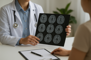
An MRI for brain stroke can show where brain tissue was injured, how large the affected area is, and whether the stroke appears recent or older. Specialized MRI sequences can detect early injury from an ischemic stroke, identify bleeding, and sometimes reveal a blocked or narrowed blood vessel. However, MRI is only one part of a stroke evaluation, and a normal scan does not always rule out a small or very early stroke.
What Can a Brain MRI Show After a Stroke?
A brain MRI uses a strong magnetic field and radio waves to create detailed images of brain tissue. It does not use ionizing radiation. The examination may include several types of images, called sequences, that provide different information.
| Possible MRI finding | What it may tell the care team |
|---|---|
| Restricted diffusion | Often indicates recent brain injury caused by interrupted blood flow. Diffusion-weighted imaging, or DWI, can detect many ischemic strokes earlier than a standard CT scan. |
| Location and size of an infarct | Shows which part of the brain was affected and may help explain problems with movement, speech, vision, balance, sensation, or other functions. |
| Blood or blood products | Blood-sensitive sequences can identify a hemorrhagic stroke, bleeding into an ischemic stroke, or evidence of previous small bleeds. |
| Swelling | Shows edema around injured tissue and whether swelling is affecting nearby brain structures. |
| Blood-vessel abnormalities | Magnetic resonance angiography, or MRA, may show a blocked, narrowed, or irregular artery. |
| Older brain injuries | May reveal previous strokes, tissue loss, scarring, or chronic changes that were not responsible for the current symptoms. |
Perfusion MRI may also evaluate how blood is moving through the brain. In selected emergency cases, perfusion information can help specialists distinguish severely injured tissue from tissue that may still be at risk. The National Institute of Neurological Disorders and Stroke overview of stroke assessment explains how MRI, CT, vascular imaging, and other tests contribute different information.
MRI Versus CT for a Suspected Stroke
Although MRI provides highly detailed images, CT is often the first scan performed in an emergency. A noncontrast head CT is fast, widely available, and effective for quickly identifying bleeding. This is important because ischemic strokes caused by blocked blood flow and hemorrhagic strokes caused by bleeding require different treatment decisions.
MRI is especially useful for detecting small ischemic strokes, strokes in the brainstem or cerebellum, and injuries that may not yet be visible on an early CT scan. Depending on the hospital, the person’s condition, and the clinical question, MRI may be performed immediately, after an initial CT, or during follow-up.
Neither scan should delay emergency evaluation. Imaging is interpreted together with the person’s symptoms, neurological examination, symptom timing, medical history, and other test results.
Can MRI Determine When a Stroke Happened?
A brain MRI after stroke can often help a radiologist classify an injury as recent, subacute, or chronic. Different sequences change in recognizable ways as the affected tissue evolves. For example, a recent ischemic injury commonly appears bright on DWI and dark on an apparent diffusion coefficient, or ADC, map. Changes on FLAIR and other sequences may become more apparent as time passes.
These patterns provide an estimate rather than an exact timestamp. The appearance can be affected by the stroke’s size, location, blood flow, treatment, and how long after symptom onset the scan was performed. Doctors therefore compare the images with the last time the person was known to be well and with any earlier scans.
What Common MRI Report Terms Mean
MRI reports contain technical language that can be concerning without context. Common terms include:
- Acute infarct: An area of recent brain injury caused by inadequate blood flow.
- Restricted diffusion: A change in water movement within tissue that strongly supports recent ischemic injury when it matches the clinical picture.
- FLAIR or T2 hyperintensity: An area that appears brighter because of increased water content or other tissue changes. It is not specific to stroke by itself.
- Hemorrhagic transformation: Bleeding that has developed within or around an area of ischemic stroke.
- Encephalomalacia or gliosis: Tissue loss or scarring from an older injury, which may have resulted from a previous stroke, trauma, or another condition.
- Chronic microvascular ischemic changes: White-matter changes often associated with aging and vascular risk factors. They are not the same as a new acute stroke.
- No acute intracranial abnormality: No new problem was identified on that examination. This does not necessarily explain ongoing symptoms or exclude every neurological condition.
The final interpretation depends on the complete report, the images themselves, and the clinical examination. MRI findings should not be used to diagnose yourself.
Can a Stroke Be Missed on MRI?
Yes. DWI is very sensitive, but it is not perfect. Very small strokes, extremely early strokes, and some strokes involving the brainstem or posterior circulation may not appear clearly on an initial scan. Movement during the examination and technical factors can also reduce image quality.
If symptoms strongly suggest a stroke despite an initially normal scan, clinicians may continue the stroke evaluation, review the images again, or order follow-up imaging. This is one reason to understand that some conditions might not show up on an MRI. A negative result should not be treated as permission to ignore new or continuing neurological symptoms.
What to Expect During the MRI
During the examination, the person lies on a table that moves into the scanner. The machine makes loud knocking or tapping sounds, so hearing protection is provided. Remaining still is important because motion can blur the images. The length of the scan varies according to the sequences needed and the person’s condition.
Many stroke MRI examinations can obtain important information without contrast. Gadolinium-based contrast may be used for certain blood-flow studies, vessel images, or when doctors are evaluating another possible cause of the symptoms. Kidney function and previous contrast reactions may be reviewed before contrast is given.
Tell the imaging team about pacemakers, aneurysm clips, cochlear implants, medication pumps, metal fragments, prior surgeries, pregnancy, kidney disease, or severe claustrophobia. Some devices are MRI-safe or MRI-conditional, while others require special precautions or may prevent scanning. The FDA’s MRI benefits and risks guidance provides additional information about magnetic-field, implant, contrast, and hearing-safety considerations.
When to Seek Care
Call 911 immediately for sudden facial drooping, weakness or numbness on one side, difficulty speaking or understanding speech, vision loss, severe loss of balance, confusion, or an unusually severe headache. Do not wait for an outpatient MRI or drive yourself to the hospital. Emergency assessment is still necessary if symptoms improve or disappear, because a transient ischemic attack can also signal an urgent risk. For more information about symptoms and stroke types, see the MedlinePlus stroke guide.
After a diagnosed stroke, report new or worsening neurological symptoms as an emergency. Questions about MRI findings can be discussed with the treating neurologist, stroke specialist, or radiologist, who can interpret the scan in combination with the examination and other testing.
Other Articles You May Find of Interest...
- How to Read a CT or MRI Report Before Your Follow-Up Appointment
- Brain MRI After a Stroke: What the Scan Can Show
- What Is Teleradiology? How It Works and What Patients Should Know
- Radiology Clinics in Southern Sydney: Options to Consider
- What Can Different Types of CT Scans Detect? A Patient Guide
- A Hypoechoic Renal Mass on Ultrasound: Next Steps
- Elastography: The Scan Replacing Some Liver Biopsies