More Imaging Articles
Elastography: The Scan Replacing Some Liver Biopsies
By Your Health Magazine Health Information Team
Your routine bloodwork shows elevated liver enzymes, and your clinician recommends checking for liver scarring. You may immediately picture a biopsy needle and days of recovery. Today, however, many people can begin with elastography—a quick, noninvasive scan that measures liver stiffness without removing tissue.
This shift is an important development in elastography news because it can make evaluating and monitoring chronic liver disease easier. Elastography does not replace every liver biopsy, but it can reduce the need for biopsy in many situations, particularly when the main question is whether significant fibrosis, or scarring, is present.
What Does Elastography Measure?
When the liver is repeatedly injured, it tries to repair itself. Over time, that healing response can create fibrous scar tissue. A liver with more fibrosis is generally stiffer than a healthy liver.
Elastography sends gentle vibrations through the liver and measures how quickly they travel. Vibrations typically move faster through stiff tissue. A computer converts those measurements into an estimate or visual map of liver stiffness.
The test is commonly used when a person has, or may be at risk for, chronic liver disease associated with:
- Metabolic dysfunction-associated steatotic liver disease, formerly called nonalcoholic fatty liver disease
- Chronic hepatitis B or hepatitis C
- Long-term heavy alcohol use
- Abnormal liver blood tests
- Other conditions that may gradually damage the liver
Fibrosis may not cause noticeable symptoms during its early stages. That makes noninvasive assessment especially useful: it may identify concerning stiffness before a person feels ill. Readers who want a broader overview can learn more about liver diseases and their possible signs and causes.
The Main Types of Liver Elastography
Ultrasound Elastography
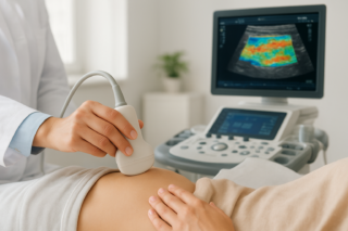
Ultrasound-based elastography is usually performed in an outpatient office or imaging center. A clinician or technologist places gel and a handheld probe against the skin on the right side of the body, often between the ribs. The device produces painless vibrations and records multiple measurements.
Vibration-controlled transient elastography is one commonly used form. Other ultrasound systems use shear-wave technology and may be incorporated into a conventional abdominal ultrasound. The elastography portion itself may take only several minutes, although the complete appointment can be longer if other imaging is performed.
Ultrasound elastography does not use ionizing radiation. Most people can return to work, drive and continue normal activities immediately afterward.
Magnetic Resonance Elastography
Magnetic resonance elastography, or MRE, combines low-frequency vibrations with MRI. A small device placed against the body creates vibrations while the scanner produces images. The resulting elastogram maps stiffness across a relatively large portion of the liver.
MRE may be helpful when ultrasound measurements are inconclusive, when more extensive liver imaging is already needed or when a specialist wants additional information. It generally takes longer, costs more and is less widely available than ultrasound elastography. People with certain implanted devices, metal in the body or severe claustrophobia may require additional screening or another approach.
For a closer look at this method, read about the benefits of MR elastography for liver health.
Why Can the Scan Replace Some Biopsies?
A liver biopsy removes a small tissue sample with a needle so a pathologist can examine it under a microscope. Biopsy can provide detailed information about fibrosis, inflammation, fat accumulation and other cellular changes. However, it is invasive and usually requires preparation, monitoring afterward and temporary limits on strenuous activity. Pain is the most common complication, while bleeding and other serious complications are less common.
Elastography does not require an incision, anesthesia or tissue removal. It also assesses a broader area of the liver than the small sample collected during a biopsy. Clinicians can repeat the scan over time with relatively little burden, making it useful for monitoring changes in chronic liver disease.
Doctors often interpret elastography together with blood tests, medical history, physical findings and standard imaging. When these pieces of information agree, they may provide enough confidence to estimate the likelihood of advanced fibrosis without a biopsy.
Does a High Stiffness Result Always Mean Scarring?
No. Increased stiffness can suggest fibrosis, but it is not a diagnosis by itself. Liver inflammation, bile-flow obstruction and congestion related to heart failure can temporarily raise stiffness. Recent food intake may also affect some measurements. Obesity, fluid in the abdomen, narrow spaces between the ribs and other technical factors can make certain ultrasound examinations difficult or less reliable.
For this reason, imaging centers may ask patients not to eat or drink for several hours before the test. Preparation instructions vary, so follow the directions from the ordering clinician or imaging facility rather than choosing a fasting period yourself.
Results may be reported as a stiffness measurement and sometimes translated into an estimated fibrosis stage. The meaning of a particular value depends on the elastography method, equipment, liver condition and quality of the readings. A number that suggests advanced fibrosis in one clinical setting should not automatically be applied to every patient or every device.
When Is a Liver Biopsy Still Necessary?
Despite promising elastography news, biopsy remains important. A clinician may recommend one when blood tests and imaging disagree, elastography results are indeterminate or the exact cause of liver injury is uncertain. Tissue may also be needed to evaluate inflammation, distinguish between overlapping liver diseases, investigate an abnormal liver lesion or confirm a diagnosis that imaging cannot establish.
Biopsy can reveal microscopic changes that stiffness measurements cannot show. For example, elastography may estimate the probability of fibrosis but cannot provide the same direct evaluation of liver cells and tissue architecture. In some conditions, it may also be better at identifying advanced scarring than separating mild, adjacent stages of fibrosis.
The practical question is therefore not whether elastography is universally better than biopsy. It is whether a noninvasive scan can answer the specific clinical question accurately enough for treatment or monitoring decisions.
What the Test May Mean for Everyday Life
For patients, elastography can turn liver monitoring into a relatively simple appointment rather than an invasive procedure. There is usually no wound care, recovery period or need to arrange transportation afterward. Repeat measurements may help the healthcare team follow trends, assess whether liver stiffness is stable and decide whether further testing is needed.
One result should not be viewed in isolation. Changes in equipment, testing conditions, inflammation and measurement quality can affect comparisons. Whenever possible, follow-up testing should be interpreted by a clinician familiar with the person’s diagnosis and previous results.
When to Seek Care
Ask a primary care clinician about liver evaluation if you have persistently abnormal liver tests, known viral hepatitis, fatty liver, heavy alcohol use or metabolic risk factors such as type 2 diabetes and obesity. A gastroenterologist or hepatologist may help determine whether blood-based fibrosis testing, ultrasound elastography, MRE or biopsy is appropriate.
Seek prompt medical care for yellowing of the skin or eyes, new abdominal swelling, vomiting blood, black or bloody stools, unusual confusion, severe weakness or significant pain in the upper-right abdomen. These symptoms do not identify a specific diagnosis, but they can occur with serious liver or digestive conditions and should not be evaluated with an elective scan alone.
Elastography represents a meaningful change in liver care: many patients can now be assessed for significant scarring without immediately undergoing biopsy. Used alongside clinical history, laboratory testing and expert interpretation, it can provide valuable information while keeping biopsy available for the situations in which tissue remains essential.
Other Articles You May Find of Interest...
- What Is Teleradiology? How It Works and What Patients Should Know
- Radiology Clinics in Southern Sydney: Options to Consider
- What Can Different Types of CT Scans Detect? A Patient Guide
- A Hypoechoic Renal Mass on Ultrasound: Next Steps
- Elastography: The Scan Replacing Some Liver Biopsies
- ‘No Suspicious Osseous Lesion’: Good News, Explained
- A Normal Pituitary MRI: What the Radiologist Saw