More Cancer Awareness Articles
Hyperplasia vs. Dysplasia: How Abnormal Cell Changes Differ
By Your Health Magazine Health Information Team
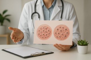
Hyperplasia means a tissue has more cells than usual, but those cells generally still look normal under a microscope. Dysplasia means the cells themselves look abnormal and may also be arranged abnormally. Neither finding automatically means cancer. However, dysplasia—especially high-grade or severe dysplasia—generally raises more concern about possible progression to cancer than simple hyperplasia does.
Hyperplasia vs. Dysplasia at a Glance
| Feature | Hyperplasia | Dysplasia |
|---|---|---|
| Basic change | Increased number of cells | Abnormal cell appearance and tissue organization |
| How cells usually look | Generally normal | Irregular in size, shape, maturity, or arrangement |
| Is it cancer? | No | No, although some forms are considered precancerous |
| Typical causes | Hormones, irritation, injury, or a normal response to increased demand | Persistent infection, chronic irritation, inflammation, or accumulated cellular changes |
| Usual description | May be physiologic, benign, or atypical | May be described as low-grade, high-grade, mild, moderate, or severe |
| Management | Depends on the organ, cause, symptoms, and presence of atypia | Often requires surveillance, additional testing, or removal of abnormal tissue |
The National Cancer Institute’s explanation of tissue changes describes hyperplasia and dysplasia as noncancerous findings that may or may not progress to cancer. They should not be viewed as automatic steps in a fixed sequence. Many hyperplastic changes never become dysplastic, and many low-grade dysplastic changes do not become cancer.
What Causes Hyperplasia?
Hyperplasia develops when cells receive signals to divide more frequently. Sometimes this is a normal and useful response. Hormonal changes, for example, stimulate growth of glandular breast tissue during pregnancy. The liver may also produce additional cells after part of it is removed or injured.
Pathologic hyperplasia occurs when growth signals are excessive or continue longer than they should. Examples include enlargement of the prostate, thickening of the uterine lining, and skin thickening caused by repeated friction. Chronic irritation and inflammation may also stimulate cell growth.
Simple hyperplasia is not necessarily precancerous. Risk depends on the specific tissue and whether the report identifies additional abnormalities. Terms such as atypical hyperplasia indicate that cells are increased in number and also look abnormal. Atypical hyperplasia of the breast or uterine lining, for example, has different implications from ordinary hyperplasia and may require closer evaluation.
What Does Dysplasia Mean?
Dysplasia describes abnormal cells within a tissue or organ. A pathologist may see differences in cell size, shape, maturity, nuclear appearance, or organization. The NCI definition of dysplasia notes that it may be classified as mild, moderate, or severe according to how abnormal the cells look and how much tissue is involved.
Dysplasia is often found in epithelial tissue—the tissue covering the skin and lining organs such as the cervix, colon, esophagus, mouth, and airways. Examples include:
- Cervical dysplasia: Frequently associated with persistent infection by certain types of human papillomavirus, or HPV.
- Colon polyp dysplasia: Abnormal cells found when an adenomatous polyp is examined after removal.
- Barrett’s esophagus with dysplasia: Abnormal changes in esophageal tissue that require specialized follow-up.
- Oral dysplasia: Abnormal cells in the lining of the mouth, sometimes associated with tobacco, alcohol, or other chronic irritation.
Dysplasia may be called low-grade when abnormalities are limited and high-grade when cells and tissue architecture are substantially altered. High-grade dysplasia generally has a greater likelihood of progressing to cancer, but the exact risk varies by organ and diagnosis.
How These Findings Differ From Cancer
Hyperplasia and dysplasia remain confined to the tissue where they developed. Cancer is defined by uncontrolled malignant growth and, in invasive cancers, the ability to grow into nearby tissue or spread elsewhere.
A report may also use terms such as carcinoma in situ or intraepithelial neoplasia. Their meaning and management vary by organ. Carcinoma in situ generally indicates highly abnormal cells that have not invaded surrounding tissue. Because terminology overlaps, the exact wording of the pathology report matters more than relying on a general definition alone.
If cancer is diagnosed, prognosis becomes a separate question influenced by the cancer type, stage, biology, age, and overall health. This can be seen in discussions of multiple myeloma prognosis across different age groups.
How Hyperplasia and Dysplasia Are Diagnosed
Imaging may identify thickened tissue, a polyp, or another abnormal area, but it usually cannot determine how individual cells look. Cytology tests, such as a Pap test, examine collected cells. A biopsy removes a tissue sample so a pathologist can evaluate both the cells and their arrangement.
As explained in this overview of biopsy procedures and laboratory analysis, samples are prepared, stained, and studied under a microscope. The pathology report may describe:
- The type of tissue sampled
- Whether hyperplasia, atypia, or dysplasia is present
- The grade or severity of abnormal changes
- Whether the sample’s edges, or margins, contain abnormal cells
- Whether there is any evidence of invasion
- Whether additional testing or a larger sample may be needed
A small biopsy represents only the area sampled. Clinicians interpret the result alongside the examination, imaging, medical history, laboratory findings, and the reason the test was performed.
What Happens After an Abnormal Finding?
Management depends on the organ involved, the cause, the degree of abnormality, and the person’s health and risk factors. Possible next steps include:
- Repeating a screening test or biopsy after a recommended interval
- Treating an underlying infection, inflammation, or hormonal imbalance
- Removing a polyp or localized abnormal area
- Using an organ-specific procedure to destroy or excise dysplastic tissue
- Having a specialist review the pathology or perform additional testing
Some mild changes resolve after the triggering factor disappears. Others persist or progress and require treatment. Follow-up schedules are specific to the diagnosis, so a cervical dysplasia plan should not be applied to dysplasia of the colon, skin, mouth, or esophagus. For additional reliable consumer resources organized by condition, visit the MedlinePlus health topics directory.
When to Seek Care
Hyperplasia and dysplasia often cause no symptoms and are discovered through screening, imaging, or biopsy. Contact the healthcare professional who ordered the test if a report mentions atypical hyperplasia, dysplasia, intraepithelial neoplasia, carcinoma in situ, unclear margins, or a recommendation for additional sampling.
Medical evaluation is also appropriate for unexplained bleeding, a persistent mouth sore, difficulty swallowing, a changing skin lesion, or another new or lasting symptom. Depending on the affected area, care may involve a primary care clinician, gynecologist, gastroenterologist, dermatologist, dentist or oral specialist, surgeon, or oncologist.
Ask what exact tissue was sampled, whether atypia is present, how the finding was graded, and what follow-up interval is recommended. Those details—not the word “hyperplasia” or “dysplasia” alone—determine what the result means for an individual patient.
Other Articles You May Find of Interest...
- Hyperplasia vs. Dysplasia: How Abnormal Cell Changes Differ
- Neck Pain Around a Chemo Port: Normal Soreness or a Complication?
- What Patients Should Know Before Starting Prostate Cancer Treatment
- Understanding CAR T Therapy: A Patient-Friendly Guide
- How Salto Health Solves The Cancer Support Gap: Why “Let Me Know If You Need Anything” Isn’t Enough
- Endocervical Adenocarcinoma: Symptoms, Diagnosis, and Treatment
- Melanoma Treatment UK: Understanding Your Care Options