




Your Health Magazine Contributor
4201 Northview Drive
Suite 102
Bowie, MD 20716
More Imaging Articles
Handheld vs Desktop Fundus Cameras: Which Is Right for Your Clinic?
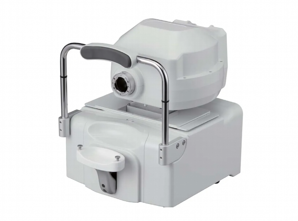
Retinal imaging has become an important part of modern eye care, helping optometrists and ophthalmologists examine, document and monitor the structures at the back of the eye. Fundus cameras capture detailed photographs of the retina, including the optic disc, macula and retinal blood vessels. These images can support clinical assessments, provide a record of retinal appearance and help practitioners identify changes that may require further investigation. As imaging technology continues to develop, eye care professionals have access to a wider range of equipment, including compact handheld devices and larger desktop systems.
Choosing between handheld and desktop fundus cameras involves more than comparing equipment size or purchase price. Image quality, patient accessibility, clinical workflow, available space and the types of examinations performed can all influence which system is most appropriate. Practices exploring fundus camera equipment should consider how different camera designs support their clinical requirements and everyday operations. Understanding the advantages and limitations of each option can help eye care professionals make informed equipment decisions while maintaining consistent standards of patient care.
Understanding the Role of Fundus Cameras in Eye Care
A fundus camera is a specialised imaging device designed to photograph the internal structures at the back of the eye. It uses an optical system and controlled illumination to capture images of the retina through the pupil. These photographs allow practitioners to document visible retinal features and assess changes associated with conditions such as diabetic retinopathy, glaucoma and age-related macular degeneration. Fundus photography can also provide useful records for comparing retinal appearance across multiple appointments.
Although retinal photographs provide valuable clinical information, they do not replace a comprehensive eye examination or other diagnostic tests. Some conditions require additional assessment using optical coherence tomography, visual field testing or other examination techniques. The usefulness of fundus photography depends on image quality, the area of the retina captured and the practitioner’s interpretation of the findings. Both handheld and desktop systems can contribute to retinal assessment, but their designs influence how images are acquired and how easily the equipment can be used in different clinical environments.
How Handheld Fundus Cameras Work
Handheld fundus cameras are compact imaging devices designed to be positioned and operated manually by a clinician. Many models incorporate digital imaging technology, integrated displays and illumination systems that allow retinal photographs to be captured without a large stationary workstation. Depending on the device, images may be viewed directly on the camera or transferred to a computer for documentation and assessment.
The main advantage of handheld cameras is their portability. Practitioners can move the equipment between examination rooms or use it in settings where a conventional desktop camera would be difficult to accommodate. This flexibility can be particularly useful when examining patients who have limited mobility or cannot comfortably position themselves at a fixed imaging station. However, handheld operation may require careful alignment and steady positioning to obtain clear photographs. The ease of use depends on the camera’s design, focusing system, image stabilisation capabilities and the operator’s experience. Some handheld models offer automated features, but the level of assistance varies considerably between devices.
Understanding Desktop and Automated Fundus Cameras
Desktop fundus cameras are generally larger systems installed in a dedicated examination area. They commonly include a stable base, adjustable patient positioning components and an integrated optical system. Patients typically sit in front of the equipment with their chin and forehead supported while the operator aligns the camera with the eye. This arrangement can help maintain a consistent imaging position during examinations.
Modern desktop systems may include automated alignment, focusing and image capture features. Some models can identify the pupil, adjust the imaging position and capture photographs with reduced manual intervention. These capabilities may improve workflow consistency, particularly in practices that perform retinal imaging frequently. However, not all desktop cameras provide the same level of automation, and some require substantial operator input. Desktop systems also require sufficient floor or bench space and appropriate patient access. Their suitability depends on the practice’s layout, examination volume and the specific imaging capabilities required rather than their size alone.
Comparing Image Quality and Imaging Capabilities
Image quality is one of the most important considerations when selecting a fundus camera. Clear retinal photographs allow practitioners to examine visible structures and compare findings across appointments. Factors affecting image quality include optical design, sensor performance, illumination, focusing accuracy and the patient’s ability to maintain a suitable position during capture.
Desktop cameras may offer advantages in positioning stability and automated image acquisition, while modern handheld devices can also produce clinically useful retinal photographs. However, image quality cannot be determined by equipment category alone. A well-designed handheld camera may outperform an older desktop model in certain circumstances, while specialised desktop equipment may provide capabilities unavailable in compact devices. Practitioners should compare specifications such as field of view, image resolution, minimum pupil size and supported imaging modes. Some systems provide colour photography only, while others offer additional imaging functions. Clinical demonstrations and sample images can help determine whether a camera meets the practice’s diagnostic and documentation requirements.
Considering Portability and Clinical Workspace
Available space can significantly influence equipment selection, particularly in smaller optometry practices or clinics with multiple consultation rooms. Desktop fundus cameras generally require a dedicated location with suitable power connections, patient seating and enough clearance for staff to operate the equipment. Once installed, they may be less convenient to move between rooms.
Handheld cameras offer greater flexibility because they can often be transported and stored more easily. This can benefit practices providing mobile eye care, visiting aged care facilities or examining patients in different clinical locations. Portable equipment may also be useful when patients cannot easily transfer to a conventional imaging station. However, portability introduces other considerations, including battery management, device protection, cleaning procedures and secure image transfer. Clinics should assess where retinal photography will take place and whether the equipment needs to be shared between practitioners. The most appropriate choice depends on how the camera will be used rather than simply whether the practice has enough space for a desktop system.
Evaluating Workflow Efficiency and Patient Needs
Clinical workflow is another important factor when comparing fundus cameras. Practices that perform a high volume of retinal imaging may benefit from equipment that supports rapid patient positioning, automated capture and straightforward image management. Desktop systems with advanced automation can help standardise certain parts of the imaging process, although actual efficiency depends on the model, operator training and patient characteristics.
Handheld cameras may provide workflow advantages in situations where transporting patients to a separate imaging room would be inconvenient. They can allow retinal photographs to be taken closer to the point of care, potentially reducing movement between clinical areas. Patient comfort should also be considered. Some individuals may find it difficult to maintain the position required by desktop equipment, while others may be easier to examine using a stable headrest and automated alignment. Patients with small pupils, cataracts or difficulty maintaining fixation may present imaging challenges regardless of the camera type. Clinics should evaluate how equipment performs with their typical patient population rather than relying exclusively on manufacturer specifications.
Choosing the Right Fundus Camera for Your Practice
Selecting a fundus camera should begin with an assessment of the practice’s clinical requirements. Practitioners should consider how frequently retinal photographs will be taken, which patient groups are commonly examined and whether imaging will occur in a fixed location or across multiple settings. It is also important to determine whether the camera needs to integrate with existing clinical software and patient record systems.
Beyond the initial purchase price, clinics should consider training, servicing, warranty coverage, software compatibility and ongoing maintenance costs. Image storage and transfer processes must also support appropriate patient privacy and information security practices. Before purchasing, practitioners may benefit from requesting a demonstration and evaluating the equipment under realistic clinical conditions. Testing image capture, patient positioning and data management can reveal practical differences that are not obvious from product descriptions. A handheld camera may be more suitable for a mobile service, while a desktop system may better support a busy fixed-location clinic. Some practices may find value in using both types for different clinical situations.
Conclusion
Handheld and desktop fundus cameras both support retinal photography, but they offer different practical advantages. Handheld systems provide portability and flexibility, making them useful for mobile services, smaller clinical spaces and patients who have difficulty accessing fixed imaging equipment. Desktop cameras may offer greater positioning stability, integrated automation and features suited to regular retinal imaging within a dedicated examination area.
The right choice depends on the clinical environment, patient population, imaging requirements and available resources. Rather than assuming that one camera type is universally superior, eye care professionals should compare individual models and assess how their features align with everyday practice needs. By considering image quality, workflow, patient accessibility and long-term equipment support, clinics can select fundus imaging technology that contributes to effective retinal documentation and informed clinical decision-making.
Other Articles You May Find of Interest...
- Handheld vs Desktop Fundus Cameras: Which Is Right for Your Clinic?
- Medical Monitor Displays for Radiology: Resolution, Calibration, and Image Quality
- Radiographer or Radiologist? Know the Difference
- What a Bone Density Test Can Tell You About Your Fracture Risk
- How to Read a CT or MRI Report Before Your Follow-Up Appointment
- Brain MRI After a Stroke: What the Scan Can Show
- What Is Teleradiology? How It Works and What Patients Should Know