More Pain Management & Rehabilitation Articles
Closed Reduction With External Fixation: How It Stabilizes a Fracture
By Your Health Magazine Health Information Team
Seeing metal bars and pins outside an injured arm or leg can be unsettling. You may wonder why a cast was not enough, whether the pins enter the bone, and how an external frame can help a fracture heal. Closed reduction external fixation combines two steps: repositioning the broken bone without surgically exposing the fracture site, then holding the corrected alignment with a stabilizing frame outside the body.
What Does Closed Reduction With External Fixation Mean?
“Reduction” means restoring the broken bone fragments to a safer, more natural position. During a closed reduction, the orthopedic surgeon adjusts the bone using traction and careful manipulation without making an incision that directly exposes the fracture.
The word “closed” describes how the bone is realigned. It does not mean that no procedure or skin incisions are involved. Small incisions are usually needed to place the fixation pins, and the procedure is commonly performed in an operating room with anesthesia. It also should not be confused with a closed fracture, in which the original injury did not break the skin.
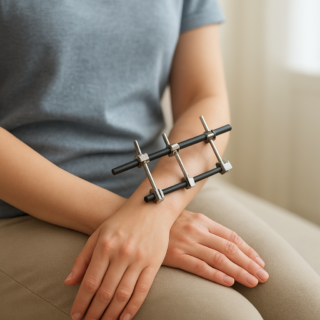
After alignment, metal pins or screws are placed through the skin into strong sections of bone above and below the break. These pins connect to rods, clamps, or rings outside the limb. In medical language, “external” simply identifies where something is located. The same word has a different meaning in conditions such as those shown in this guide to real images of external hemorrhoids.
How the External Fixator Stabilizes the Fracture
A displaced or unstable fracture can shorten, rotate, or shift because of muscle pull, gravity, swelling, and normal movement. The external fixator acts as a rigid bridge around the injured area. Once its pins and connecting bars are secured, the device helps maintain the bone’s length, alignment, and rotation.
This stability limits unwanted movement at the fracture while the body begins producing new bone. Depending on the injury, the frame may also span a nearby joint to keep the entire region still. Because much of the device remains outside the body, clinicians can continue examining wounds and swollen soft tissues around the fracture.
External fixation may be temporary or definitive. A temporary frame can hold the bone safely while swelling decreases, wounds receive treatment, or the patient recovers from other serious injuries. The surgeon may later replace it with plates, screws, or a rod. In selected cases, the external fixator remains in place until the fracture has healed sufficiently.
When Is This Treatment Used?
Closed reduction with external fixation is generally considered when a fracture needs more support than a cast or splint can provide, but opening the fracture site or immediately placing internal hardware may create additional risk. Common situations include:
- Unstable or severely displaced fractures
- Fractures broken into several pieces
- Major swelling, blistering, or soft-tissue damage
- Open fractures with wounds requiring ongoing access
- Complex fractures near the ankle, knee, wrist, or elbow
- Multiple traumatic injuries requiring rapid stabilization
- Temporary stabilization before internal fixation
- Selected fractures for which external fixation is the planned final treatment
The decision depends on the fracture pattern, condition of the skin and muscles, circulation and nerve function, infection risk, overall health, and expected ability to follow care instructions.
What Happens During the Procedure?
Before treatment, the clinical team examines circulation, sensation, movement, and the surrounding skin. X-rays and sometimes other imaging help define the fracture. Pain control may involve regional anesthesia, sedation, or general anesthesia, depending on the injury and procedure.
The surgeon then applies controlled pulling and manipulation to restore alignment. Live X-ray imaging may be used to check the bone position. Pins or screws are inserted into uninjured bone at a safe distance from the fracture and connected to the external frame. The surgeon tightens the frame after confirming the desired length and alignment, and additional images are obtained before the procedure ends.
The fixator does not “glue” the bone together or make healing immediate. It creates the mechanical conditions needed for healing while the body repairs the fracture over time.
Living With an External Fixator
A frame can make dressing, sleeping, bathing, and moving around more complicated. Loose clothing may fit more comfortably, while pillows may help support the limb without pressing on the device. Patients should follow their orthopedic team’s instructions about weight-bearing, elevation, showering, physical therapy, and activity. Weight-bearing rules vary widely; the presence of a sturdy frame does not automatically mean it is safe to walk on the injured limb.
Pin sites require close observation because each pin passes through the skin. There is no single care routine appropriate for every frame, so patients should use only the cleaning and dressing method provided by their surgical team. The frame should not be adjusted, tightened, covered with unapproved materials, or used as a handle.
Follow-up visits and imaging are essential. The surgeon checks alignment, bone healing, pin stability, wounds, and joint movement. Physical or occupational therapy may help preserve strength and motion in areas that can safely move. Broader educational information about fractures and other conditions is available through the MedlinePlus health topics library.
Possible Risks and Complications
Every fracture and fixation procedure has risks. With external fixation, the most frequent concern is irritation or infection where a pin enters the skin. Warning signs can include increasing redness, warmth, swelling, tenderness, drainage, or pain around a pin.
Other possible complications include pin loosening, frame failure, loss of fracture alignment, stiffness, nerve or blood-vessel injury, delayed healing, failure of the bone to unite, or healing in an incorrect position. Prolonged immobility can also contribute to muscle weakness and blood-clot risk. Regular monitoring allows the orthopedic team to identify many problems before they significantly affect treatment.
When to Seek Care
Contact the orthopedic surgeon promptly for worsening pin-site redness, new drainage, fever, increasing pain, a loose pin, a bent or damaged frame, or an unexpected change in the limb’s position. Urgent evaluation is appropriate for severe or rapidly increasing pain, major swelling, new numbness or weakness, or fingers or toes that become pale, blue, or cold. Chest pain, sudden shortness of breath, or coughing blood requires emergency medical attention.
Closed reduction external fixation can look intimidating, but its purpose is straightforward: restore alignment without opening the fracture site and maintain that position with a strong external support. For carefully selected injuries, it protects damaged tissues, provides access for wound care, and creates a stable environment in which bone healing can begin.
Other Articles You May Find of Interest...
- What to Do If You Think You Have a Broken Tailbone
- Closed Reduction With External Fixation: How It Stabilizes a Fracture
- Knee Massager for Muscle Tension Around the Knee What You Need to Know
- What Life Really Looks Like a Year After Spine Surgery
- Gout or Bursitis? Why Joint Swelling Can Look the Same
- Broken Kneecap Recovery: How Long Healing Usually Takes
- The Thumb Grind Test: What Pain During It Can Mean