


Your Health Magazine
4201 Northview Drive
Suite #102
Bowie, MD 20716
301-805-6805
More Health News & Research Articles
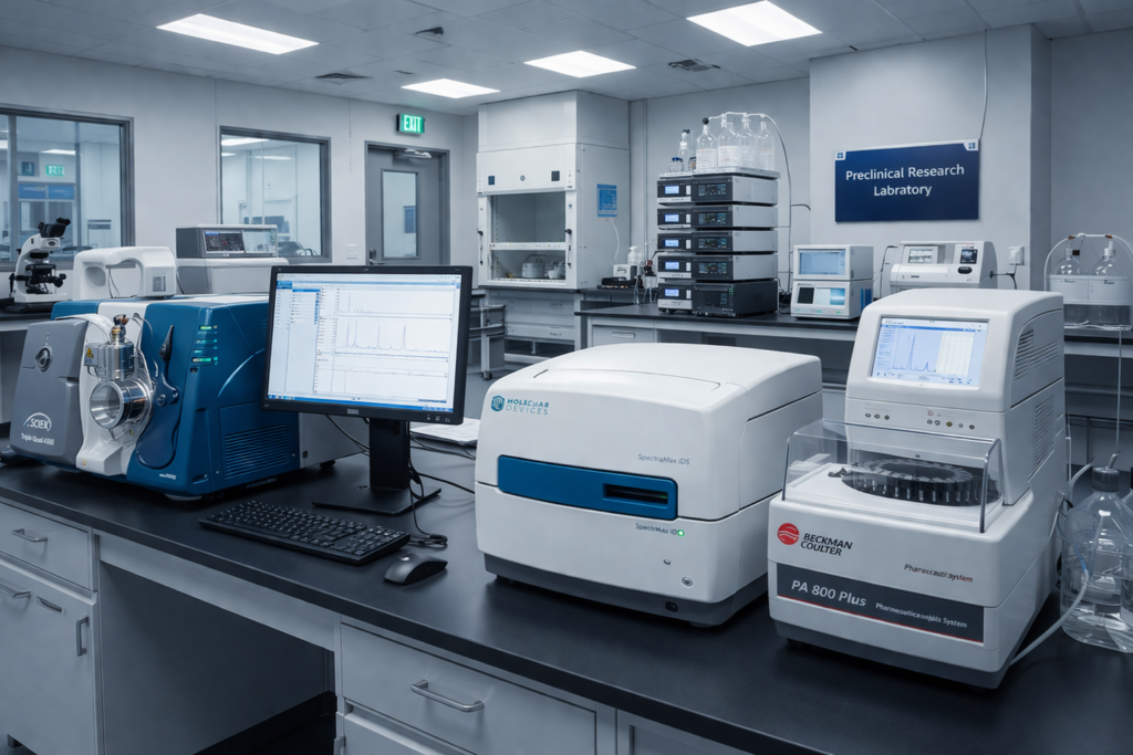
How Researchers Measure Intracellular Glutathione Levels In Preclinical Studies
Researchers measure intracellular glutathione levels in preclinical studies using enzymatic recycling assays, HPLC with mass spectrometry, fluorescent probe-based assays, genetically encoded biosensors, and capillary electrophoresis. Each method quantifies reduced glutathione (GSH), oxidized glutathione (GSSG), or both within cell lysates, tissue homogenates, and live-cell models. The enzymatic recycling assay (DTNB/Ellman’s method) is the most widely adopted technique for total glutathione measurement due to its accessibility and reproducibility. HPLC-MS/MS provides the highest specificity for simultaneous GSH and GSSG resolution. Genetically encoded biosensors such as roGFP2-Grx1 enable real-time, non-destructive monitoring of the glutathione redox potential at the subcellular level. Selecting the right analytical approach depends on the research objective, required sensitivity, sample type, and throughput demands of the preclinical study design.
Glutathione is the most abundant low-molecular-weight thiol in mammalian cells and serves as a primary regulator of intracellular redox homeostasis. Accurate GSH/GSSG ratio measurement is foundational to oxidative stress research, toxicology screening, and mechanistic studies of cellular antioxidant defense pathways. This article breaks down each method, compares their strengths and limitations, and outlines best practices for sample handling that prevent artifactual oxidation from compromising data integrity.
Disclaimer: Glutathione is sold strictly for research purposes only and is not intended for human consumption. The following content is provided for educational and informational purposes to support professional researchers and academics. Nothing in this article constitutes medical advice, a therapeutic recommendation, or an endorsement of any compound for clinical use. All references to glutathione pertain exclusively to in vitro and preclinical laboratory applications.
Why Intracellular Glutathione Measurement Matters in Research
Glutathione is the most abundant low-molecular-weight thiol in mammalian cells. It functions as a critical component of the cellular antioxidant defense system, participating in detoxification reactions, protein folding, and redox homeostasis. In preclinical research, measuring GSH levels helps investigators understand how cells respond to environmental stressors, toxic exposures, and experimental interventions.
The ratio of reduced glutathione (GSH) to oxidized glutathione (GSSG) serves as a widely recognized indicator of intracellular redox status. A declining GSH/GSSG ratio in cell culture or tissue samples may signal increased oxidative burden, making this measurement essential in toxicology studies, pharmacokinetic research, and mechanistic investigations of cellular pathology.
Researchers also monitor glutathione levels to evaluate the activity of enzymes such as glutathione peroxidase (GPx), glutathione S-transferase (GST), and glutathione reductase (GR). These enzymes are central to phase II detoxification and reactive oxygen species (ROS) scavenging, both of which are active areas of preclinical investigation.
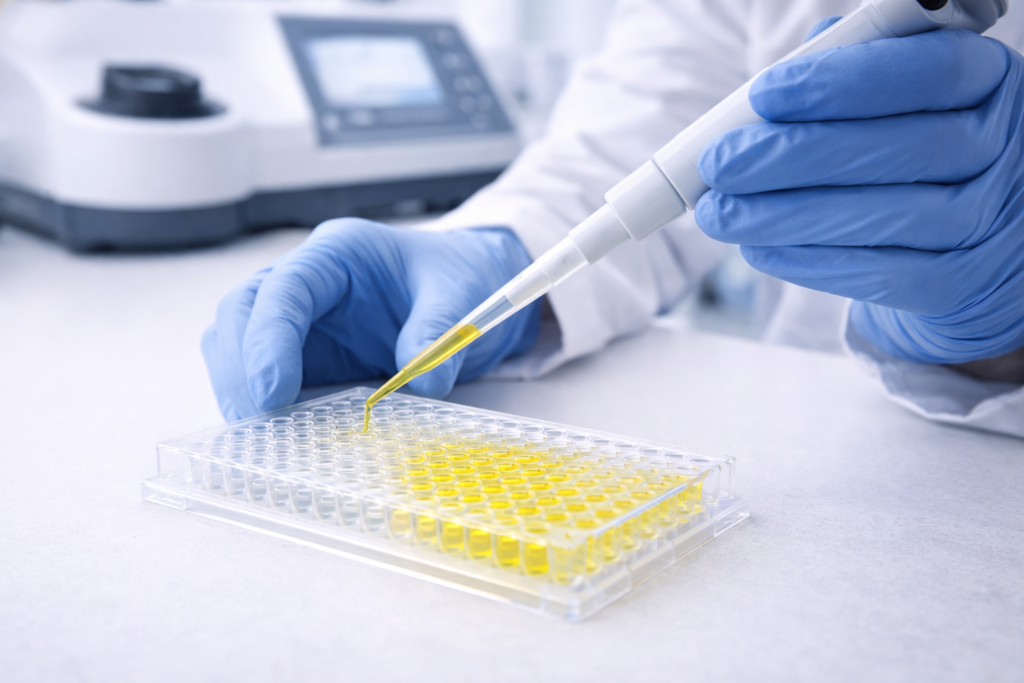
Enzymatic Recycling Assay (Ellman’s Method / DTNB Assay)
Overview
The enzymatic recycling assay, often referred to as the DTNB assay or Ellman’s method, is one of the most established techniques for total glutathione quantification in research laboratories. This colorimetric approach relies on the reaction between 5,5′-dithio-bis(2-nitrobenzoic acid) (DTNB) and the sulfhydryl group of GSH.
How It Works
In this assay, DTNB reacts with GSH to produce a mixed disulfide and the chromophore 5-thio-2-nitrobenzoic acid (TNB). The enzyme glutathione reductase then reduces the mixed disulfide back to GSH using NADPH as a cofactor. This recycling mechanism amplifies the signal, allowing researchers to detect total glutathione (GSH + GSSG) with high sensitivity.
To measure GSSG specifically, researchers first derivatize free GSH using 2-vinylpyridine (2-VP) or N-ethylmaleimide (NEM). This blocks the reduced thiol and prevents it from participating in the recycling reaction, isolating GSSG for quantification.
Advantages in Preclinical Applications
The enzymatic recycling assay offers several benefits for laboratory research. It is relatively inexpensive, uses standard spectrophotometric equipment, and provides reproducible results across a range of biological matrices including cell lysates, tissue homogenates, and plasma samples. Its linear dynamic range typically spans from low micromolar to millimolar concentrations, making it suitable for most preclinical sample types.
Limitations
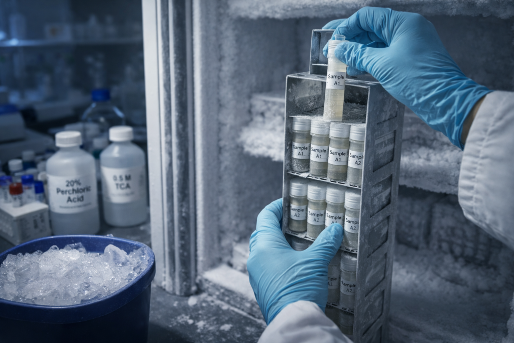
This method measures total glutathione rather than providing spatial or subcellular resolution. It also requires careful sample preparation to prevent artifactual oxidation of GSH during extraction. Researchers working with this assay commonly use metaphosphoric acid (MPA) or perchloric acid (PCA) to deproteinize samples immediately upon collection.
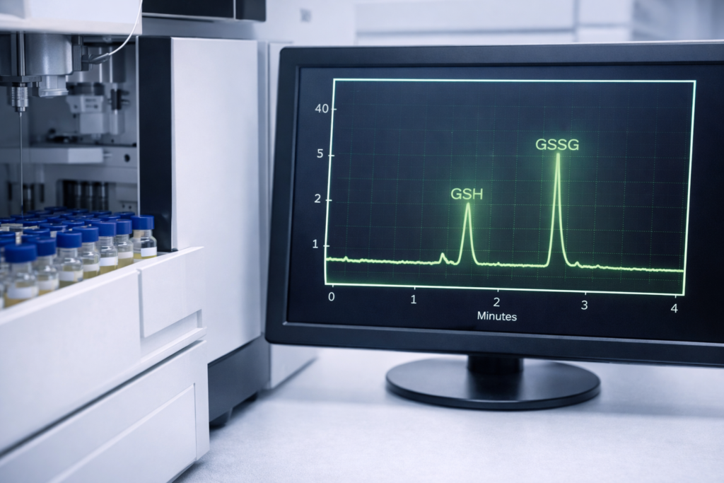
High-Performance Liquid Chromatography (HPLC)
Overview
HPLC-based methods remain the gold standard for simultaneous quantification of GSH and GSSG in preclinical research. These techniques offer excellent specificity, sensitivity, and the ability to resolve glutathione from other thiol-containing compounds in complex biological samples.
Detection Approaches
Researchers use several detection modalities paired with HPLC separation:
Electrochemical detection (ECD): This approach measures the oxidation or reduction current generated by glutathione at an electrode surface. HPLC-ECD is highly sensitive and can detect picomole quantities of GSH and GSSG, making it ideal for small tissue samples or limited cell populations.
Ultraviolet (UV) detection: Following derivatization with reagents such as iodoacetic acid and 1-fluoro-2,4-dinitrobenzene (FDNB), glutathione species absorb UV light at characteristic wavelengths. UV detection provides reliable quantification but typically requires larger sample volumes compared to ECD.
Fluorescence detection: Derivatization with fluorescent tags such as monobromobimane (mBBr) or o-phthalaldehyde (OPA) enables highly sensitive fluorescence detection. This method is particularly useful when researchers need to quantify glutathione in samples with low analyte concentrations.
Mass spectrometry (MS): LC-MS and LC-MS/MS provide the highest specificity and sensitivity for glutathione analysis. These techniques can differentiate glutathione from structurally similar compounds and quantify multiple thiol species in a single analytical run. Isotope-dilution mass spectrometry further improves accuracy by correcting for matrix effects.
Sample Preparation Considerations
Proper sample handling is critical for HPLC-based glutathione analysis. Rapid acid precipitation or alkylation of free thiols immediately after sample collection prevents ex vivo oxidation. Many researchers add internal standards at the point of extraction to account for recovery losses throughout the analytical workflow.
Fluorescent and Luminescent Probe-Based Assays
Overview
Fluorescent probe-based methods have become increasingly popular in glutathione research due to their compatibility with high-throughput screening, live-cell imaging, and microplate reader formats. These assays use synthetic probes that react selectively with glutathione to generate a fluorescent or luminescent signal proportional to analyte concentration.
Commonly Used Probes
Monochlorobimane (mCB): This cell-permeable probe reacts with GSH in a reaction catalyzed by glutathione S-transferase, producing a fluorescent GSH-bimane conjugate. Researchers frequently use mCB for live-cell imaging of glutathione distribution using confocal microscopy or flow cytometry.
ThiolTracker Violet: This commercially available probe provides a bright, photostable fluorescent signal upon reaction with intracellular thiols, predominantly GSH. It is compatible with both microscopy and plate-based quantification.
Naphthalimide-based probes: Several research groups have developed naphthalimide derivatives that exhibit turn-on fluorescence upon reaction with GSH. These probes offer tunable excitation and emission wavelengths, enabling multiplexed imaging alongside other fluorescent markers.
Luciferin-based luminescent assays: Bioluminescent approaches use modified luciferin substrates that release free luciferin upon reaction with GSH. The resulting luminescent signal, measured with a standard luminometer, provides excellent signal-to-noise ratios and minimal background interference.
Applications in High-Throughput Research
Probe-based assays are particularly valuable in preclinical screening studies where researchers need to evaluate glutathione modulation across hundreds or thousands of experimental conditions. Microplate-compatible formats allow rapid data acquisition, and many probes are compatible with automated liquid handling systems used in academic and contract research laboratories.
Considerations for Probe Selection
Researchers should evaluate probe specificity carefully. Some fluorescent indicators react with cysteine, homocysteine, or other biological thiols in addition to glutathione. Selectivity validation using GSH-depleted controls (e.g., buthionine sulfoximine [BSO]-treated cells) is a standard practice in well-designed preclinical studies.
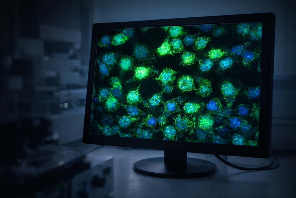
Genetically Encoded Biosensors
Overview
Genetically encoded glutathione biosensors represent a significant advancement in redox biology research. These protein-based tools allow real-time, reversible monitoring of the glutathione redox potential (E_GSH) within living cells, offering subcellular resolution that chemical probes cannot match.
Key Biosensor Platforms
roGFP2-Grx1: This widely used biosensor consists of a redox-sensitive green fluorescent protein (roGFP2) fused to human glutaredoxin-1 (Grx1). The Grx1 domain equilibrates rapidly with the local GSH/GSSG pool, transferring the redox information to roGFP2. The resulting ratiometric fluorescence change (excitation at ~400 nm and ~490 nm) provides a quantitative readout of the glutathione redox potential.
rxYFP: Redox-sensitive yellow fluorescent protein variants respond to changes in the thiol-disulfide equilibrium. While less specific to glutathione than Grx1-coupled sensors, rxYFP constructs have been used in compartment-specific redox measurements in preclinical models.
Subcellular Targeting
A major advantage of genetically encoded sensors is the ability to target them to specific organelles. Researchers have expressed roGFP2-Grx1 in the cytosol, mitochondrial matrix, endoplasmic reticulum, and nucleus to map compartmentalized glutathione redox dynamics. This spatial resolution is critical for understanding how oxidative stress affects distinct cellular compartments in preclinical disease models.
Limitations
Genetically encoded biosensors require transfection or viral transduction, which may not be feasible in all experimental systems. They also measure the glutathione redox potential rather than absolute GSH concentration, which is an important distinction researchers must account for when interpreting results.
Capillary Electrophoresis (CE)
Capillary electrophoresis offers an alternative separation technique for glutathione analysis in preclinical samples. CE methods provide rapid separation times, low sample volume requirements, and compatibility with UV, fluorescence, and mass spectrometric detection.
CE coupled with laser-induced fluorescence (CE-LIF) detection achieves exceptionally low detection limits for derivatized glutathione species. This approach is particularly useful for single-cell analysis, where researchers need to quantify glutathione in individual cells isolated from heterogeneous populations.
The small sample requirements of CE-based methods make them attractive for preclinical studies involving limited tissue availability, such as microdissected brain regions or small biopsy specimens from animal models.
Spectrophotometric and Colorimetric Kits
Commercial glutathione assay kits are widely available and provide standardized protocols for laboratories that need reliable, reproducible results without extensive method development. These kits typically use variations of the enzymatic recycling assay or proprietary chromogenic substrates.
Kit-based assays offer convenience and batch-to-batch consistency, which is valuable for multi-site preclinical studies requiring harmonized analytical methods. Many kits include pre-optimized reagents, calibration standards, and detailed protocols that reduce inter-operator variability.
Researchers should verify kit performance in their specific sample matrix, as components in cell lysates or tissue homogenates can interfere with colorimetric reactions. Running spike-and-recovery experiments during method validation ensures accurate quantification in the intended biological context.
Comparing Methods: Selecting the Right Approach
Choosing an appropriate glutathione measurement method depends on the specific research question. Below is a summary of key considerations:
For total glutathione quantification in bulk samples: The enzymatic recycling assay or HPLC with electrochemical detection provides reliable results with well-established protocols.
For simultaneous GSH and GSSG measurement: HPLC-MS/MS or HPLC with fluorescence detection offers the specificity needed to resolve both species in a single run.
For live-cell imaging and spatial resolution: Genetically encoded biosensors such as roGFP2-Grx1 or cell-permeable fluorescent probes like monochlorobimane are the preferred tools.
For high-throughput screening: Microplate-compatible fluorescent or luminescent assays allow rapid evaluation across large experimental panels.
For single-cell analysis: Capillary electrophoresis with laser-induced fluorescence detection provides the sensitivity and resolution required for individual cell measurements.
Best Practices for Glutathione Sample Handling in the Laboratory
Regardless of the analytical method selected, proper sample handling is essential for accurate glutathione measurement. Artifactual oxidation of GSH to GSSG during sample processing is a well-documented source of error in preclinical studies.
Researchers should adhere to the following practices to preserve sample integrity. Rapid deproteinization using acidic solutions (MPA, PCA, or trichloroacetic acid) immediately upon sample collection prevents enzymatic degradation and spontaneous oxidation. Alkylating agents such as NEM or iodoacetamide can be added to block free thiols and lock the GSH/GSSG ratio at the time of collection. Samples should be processed on ice and stored at minus 80 degrees Celsius until analysis. Repeated freeze-thaw cycles should be avoided, as they promote oxidation and reduce measurement accuracy.
Inclusion of appropriate quality control measures, including method blanks, spiked recovery samples, and certified reference materials, strengthens the reliability of glutathione data in any preclinical study.
Emerging Technologies in Glutathione Research
The field of glutathione measurement continues to advance. Several emerging approaches are gaining traction in preclinical research settings:
Raman spectroscopy: Surface-enhanced Raman spectroscopy (SERS) enables label-free detection of glutathione in biological samples. Researchers are developing SERS-based platforms for rapid, non-destructive glutathione analysis in cell cultures and tissue sections.
Electrochemical biosensors: Miniaturized electrochemical sensors incorporating glutathione-responsive nanomaterials (gold nanoparticles, carbon nanotubes, graphene oxide) offer the potential for continuous, real-time glutathione monitoring in perfused tissue models and organ-on-a-chip systems.
Mass spectrometry imaging (MSI): Matrix-assisted laser desorption/ionization (MALDI) MSI allows researchers to map glutathione distribution across tissue sections with spatial resolution on the order of tens of micrometers. This technique provides both quantitative and spatial information in a single experiment.
These technologies are still under active development, but they represent promising tools for the next generation of preclinical redox biology research.
Conclusion
Selecting the right glutathione measurement method is a critical decision that shapes the quality and reliability of preclinical research outcomes. Researchers should match their analytical approach to the specific demands of their study, whether that means deploying HPLC-MS/MS for precise GSH/GSSG quantification, using genetically encoded biosensors like roGFP2-Grx1 for real-time subcellular redox mapping, or leveraging high-throughput fluorescent assays for large-scale screening campaigns. Every method requires disciplined sample handling, including immediate deproteinization, thiol alkylation, and strict temperature control, to prevent artifactual oxidation from compromising data integrity. As emerging platforms such as SERS-based sensors, electrochemical nanomaterial arrays, and MALDI mass spectrometry imaging continue to mature, researchers will gain even greater precision in mapping intracellular glutathione dynamics across complex biological systems. Investing in proper method validation and quality control protocols today positions preclinical laboratories to generate reproducible, publication-ready glutathione data that advances the broader understanding of redox biology.
FAQs
What is the most widely used method for measuring total glutathione in preclinical research?
The enzymatic recycling assay (DTNB/Ellman’s method) remains the most widely adopted technique for total glutathione quantification in research laboratories. It uses glutathione reductase and NADPH to amplify a colorimetric signal that standard spectrophotometers can detect with high reproducibility. Researchers favor this approach for its accessibility, well-documented protocols, and compatibility with diverse biological sample types including cell lysates and tissue homogenates.
How do researchers distinguish between reduced glutathione (GSH) and oxidized glutathione (GSSG)?
Researchers block free GSH using alkylating agents such as 2-vinylpyridine (2-VP) or N-ethylmaleimide (NEM) before running the assay, which isolates GSSG for independent measurement. HPLC paired with mass spectrometry or fluorescence detection can also resolve both species simultaneously within a single analytical run. Calculating the GSH/GSSG ratio from these measurements provides a reliable indicator of intracellular redox status in preclinical models.
Why is sample handling so important when measuring intracellular glutathione levels?
Glutathione is highly susceptible to artifactual oxidation during sample collection, processing, and storage, which can significantly skew GSH/GSSG ratios. Researchers prevent this by immediately deproteinizing samples with acidic solutions such as metaphosphoric acid (MPA) or perchloric acid (PCA) and processing all materials on ice. Avoiding repeated freeze-thaw cycles and storing samples at minus 80 degrees Celsius further preserves the native redox state of the analyte.
Can researchers measure glutathione in live cells without destroying the sample?
Yes. Genetically encoded biosensors such as roGFP2-Grx1 allow real-time, reversible monitoring of the glutathione redox potential inside living cells without requiring lysis or fixation. Cell-permeable fluorescent probes like monochlorobimane (mCB) and ThiolTracker Violet also enable live-cell glutathione imaging using confocal microscopy or flow cytometry platforms.
What analytical method is best suited for high-throughput glutathione screening in preclinical studies?
Microplate-compatible fluorescent and luminescent probe-based assays are the preferred choice for high-throughput glutathione screening across large experimental panels. These formats integrate seamlessly with automated liquid handling systems and standard plate readers found in most academic and contract research laboratories. Researchers should validate probe specificity using GSH-depleted controls, such as buthionine sulfoximine (BSO)-treated cells, to confirm that the signal reflects glutathione rather than other intracellular thiols.
Other Articles You May Find of Interest...
- Pooled Human Hepatocytes: Improving Drug Metabolism Studies with Diverse Donor Profiles
- How Researchers Measure Intracellular Glutathione Levels In Preclinical Studies
- Chemistry Reagents: What They Are, What They Do, and Why How You Handle Them Decides Everything
- A Practical Peptide Selection Guide For Laboratory Buyers
- What Is Cardarine (GW-501516)? A Preclinical Research Overview
- Troubleshooting Common ELISA Immunoassay Kit Errors
- Best Practices for Choosing USA Made Research Peptides


