


More Vascular Health Articles
Unraveling the Secrets of Pulmonary Embolism ECG: What the Readings Reveal
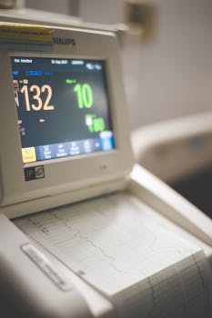
When it comes to understanding the complexities of heart health, the pulmonary embolism ECG plays a vital role in diagnosing medical emergencies. A pulmonary embolism occurs when a blood clot gets lodged in an artery in the lungs, blocking blood flow. As this condition can be life-threatening, early detection and efficient diagnosis using tools like an ECG can potentially save lives.
Understanding Pulmonary Embolism ECG
An ECG, or electrocardiogram, is a practical and fast tool health professionals use to check the heart’s electrical activity. While it cannot definitively diagnose a pulmonary embolism, it can reveal patterns that indicate its presence. Changes in the ECG might prompt further testing like CT scans or D-Dimer blood tests to confirm the diagnosis.
The ECG’s primary capability is to detect abnormalities in heart functions that may suggest a pulmonary embolism. Signs like sinus tachycardia, right ventricular strain patterns, or the classic S1Q3T3 pattern are typically monitored. However, these readings aren’t exclusive to embolisms and must be interpreted by a clinician in context with other symptoms and tests.
Common Indicators and Patterns
While the ECG is a great tool, it is worth stating that symptoms and results vary among individuals. Here are some common patterns associated with a pulmonary embolism ECG:
- Sinus Tachycardia: This is the most common complication found on an ECG, reflecting the heart’s increased rate as it tries to maintain circulation despite blockages.
- Right Bundle Branch Block (RBBB): This indicates an obstruction in the electrical signals of the right side of the heart, which is significant in the presence of acute pulmonary conditions.
- S1Q3T3 Pattern: Although not uniquely indicative of a pulmonary embolism, this pattern is often checked. It involves large S waves in lead I, a Q wave in lead III, and an inverted T wave in lead III.
Recognizing these patterns helps guide medical professionals towards an accurate diagnosis. Doctors may also look for right axis deviation or T-wave inversions in the anterior leads to support their findings.
Beyond ECG Readings
It’s important to understand that an ECG is only one part of the diagnostic puzzle. As pulmonary embolisms require timely treatment, healthcare practitioners might order further imaging or blood tests to confirm the presence of a clot. These follow-up procedures might include a CT pulmonary angiogram or a D-dimer test, which is a blood test that measures clot formation and breakdown in the body.
When an ECG Might Be Needed
Individuals showing symptoms such as sudden shortness of breath, chest pain, or a rapid heart rate may need a pulmonary embolism ECG to assess heart function. However, changes in lifestyle choices can influence overall cardiovascular health and potentially alter these symptoms. Understanding how lifestyle adjustments affect heart health can be beneficial in preventing such emergencies.
Consulting Healthcare Professionals
It is crucial to consult with healthcare providers if you suspect a pulmonary embolism or if new symptoms arise. Since each patient’s condition is unique, a personalized approach to care is essential. A consultation ensures that any interventions complement the patient’s overall health context.
The Role of ECGs in Diagnosing Pulmonary Embolism
While ECGs are a non-invasive and quick method to garner initial insights, their utility lies in conjunction with other diagnostic tests. The ECG identifies abnormal heart rhythms and other signals indicative of potential pulmonary embolisms, offering valuable information for determining the next steps for care.
For more detailed and scientifically-backed information on ECGs and pulmonary health, you could explore the health section of Wikipedia as a starting point.
In conclusion, the pulmonary embolism ECG is an essential tool that assists in the initial identification of potential pulmonary blockages, though further tests are often needed for confirmation. Understanding these readings and the symptoms related to pulmonary embolism enhances preparedness and response in medical settings.
- ECG changes can suggest pulmonary embolism but are not definitive.
- Common patterns include sinus tachycardia and right bundle branch block.
- Additional tests like CT scans are necessary for confirmation.
- An integrated approach to care includes lifestyle considerations.
FAQs
What are the primary signs of a pulmonary embolism on an ECG?
While various patterns can appear, sinus tachycardia and the S1Q3T3 pattern are among the most observed. These patterns necessitate further medical evaluation.
Why is an ECG not enough to diagnose a pulmonary embolism?
An ECG can show indirect signs of stress or blockage but lacks the specificity needed, requiring additional imaging or blood tests for accurate diagnosis.
How quickly should a pulmonary embolism be treated?
A pulmonary embolism needs immediate attention due to its potential severity. Prompt medical care reduces risk and improves outcomes.
Can lifestyle changes reduce the risk of a pulmonary embolism?
Yes, maintaining a healthy lifestyle with regular exercise, a balanced diet, and avoiding smoking can contribute to reducing the risk.
Is the ECG used in isolation for diagnosing heart-related issues?
No, the ECG is typically used alongside other diagnostic tools to provide a comprehensive view of heart health, especially in the presence of concerning symptoms.
Other Articles You May Find of Interest...
- The Timeline of Imiquimod: When Can You Expect Results?
- Discover the Benefits and Risks of the Strongest Blood Thinner
- Exploring the Benefits and Uses of Isosorbide Mononitrate in Heart Health
- Navigating Life with a Transvenous Pacemaker: Essential Insights for Patients
- Is the Trendelenburg Sign a Cause for Concern?
- Essential Insights About the Quinton Catheter for Effective Health Management
- Bevacizumab Side Effects: What Patients Should Be Aware Of


