


More Heart Disease, Stroke and Diabetes Articles
Unraveling the Mysteries of ECG T Wave Abnormalities
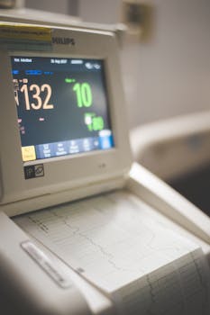
Understanding ECG T wave abnormalities can seem daunting, but it is a crucial factor in comprehending heart health. An ECG, or electrocardiogram, records the electrical activity of the heart over time. The T wave is a critical component of this reading, representing the heart’s electrical recovery phase. When an abnormality is detected in the T wave, it may indicate underlying heart issues. This article aims to explore what an ECG T wave abnormality entails, its potential causes, and what they might mean for your health.
What is an ECG T Wave Abnormality?
An ECG T wave abnormality occurs when the shape, size, or pattern of the T wave deviates from the norm. This can happen for a variety of reasons, some benign and others indicative of a more serious condition. Understanding these variations is important for interpreting the potential implications for heart health, as a wide range of heart diseases can alter the T wave.
Common Types and Causes of T Wave Abnormalities
T wave abnormalities can manifest in several ways, including flattened, inverted, or peaked T waves. Each pattern may point to different underlying issues:
- Flattened T waves: These may suggest electrolyte imbalances, such as low potassium levels.
- Inverted T waves: Commonly associated with ischemia, where there is reduced blood flow to the heart.
- Peaked T waves: Often linked with hyperkalemia, where high potassium levels affect heart function.
While these are common examples, it’s important to remember that ECG readings must be interpreted in the context of a full clinical picture.
When Should You Be Concerned?
While an ECG T wave abnormality can sometimes be harmless, there are instances when it warrants further investigation. For example, if the abnormality is associated with symptoms such as chest pain, dizziness, or shortness of breath, medical advice should be sought immediately. Additionally, individuals with a history of heart conditions, such as hypertension or prior heart attacks, should be more vigilant.
Diagnosing ECG T Wave Abnormalities
Diagnosis of an ECG T wave abnormality typically involves a detailed review of the patient’s history, symptoms, and other diagnostic tests. A physician may conduct a stress test or an echocardiogram for further insight. Lifestyle factors, such as diet and exercise, can also play a role in heart health. To learn more about this, consider reading our article on how lifestyle choices impact our overall health.
Implications for Treatment
The treatment of ECG T wave abnormalities depends heavily on the underlying cause. In some cases, managing existing health conditions can resolve the abnormality. Medications, lifestyle changes, and regular monitoring might be recommended. It is crucial to have an ongoing discussion with a healthcare provider to adjust treatment plans as needed.
Conclusion
Navigating ECG T wave abnormalities involves understanding their potential implications and seeking appropriate medical guidance. While not all abnormalities indicate significant issues, they require careful consideration. An ECG T wave abnormality should never be ignored, especially if accompanied by troubling symptoms. For more information about heart health and medical conditions, you can refer to trusted resources such as the World Health Organization’s health section.
- ECG T wave abnormalities can be caused by various factors, including electrolyte imbalances and ischemia.
- Diagnosis involves considering clinical history and additional tests.
- Symptoms accompanying T wave abnormalities, like chest pain, should prompt immediate medical attention.
- Management may include lifestyle changes, medication, or monitoring.
- Consulting a clinician is crucial for personalized guidance.
What is an ECG?
An ECG, or electrocardiogram, is a test that measures the electrical activity of the heart through electrodes placed on the skin. Each heartbeat is represented by waves, including the P wave, QRS complex, and T wave.
What does an inverted T wave indicate?
An inverted T wave on an ECG may indicate conditions such as myocardial ischemia, where blood flow to the heart is reduced, or it might be a normal variant. It is essential to correlate ECG findings with clinical evaluation.
Can anxiety affect the ECG results?
Yes, anxiety and other stress-related conditions can affect heart rate and rhythm, potentially leading to transient changes in ECG readings. It’s important to inform your healthcare provider if you’re feeling anxious during the test.
What are the risk factors for T wave abnormalities?
Risk factors include a history of heart disease, high blood pressure, electrolyte imbalances, and certain medications. Managing these risks through lifestyle choices and medical care is essential.
How are electrolyte imbalances detected?
Electrolyte imbalances can be detected through blood tests that measure levels of key electrolytes, such as potassium, sodium, and magnesium. These tests help identify possible causes of ECG changes.
Other Articles You May Find of Interest...
- How Long Do Blood Pressure Monitors Last?
- Optimal Timing for Taking Januvia to Maximize Its Benefits
- Does Rosuvastatin Act as a Blood Thinner? Discover the Facts
- Is Januvia a Hypoglycemic Drug That Can Help Manage Diabetes?
- How to Prevent Heart Attacks Before They Happen
- The Impact of Furosemide on Blood Pressure: What You Need to Know
- Is Your Chest Pain Accompanied by a Metallic Taste in Your Mouth?


