


More Imaging Articles
Exploring the Role of Isoechoic Structures in Medical Imaging
In the realm of medical imaging, particularly in ultrasound technology, understanding the concept of isoechoic structures is crucial. These structures display echo patterns similar to surrounding tissues, making them challenging to identify without a trained eye. For patients and healthcare providers alike, deciphering the subtleties of isoechoic patterns is key to accurate diagnosis and treatment planning.
Understanding Isoechoic Structures
When a tissue or mass is termed isoechoic, it means that it produces ultrasound echoes of the same intensity as the surrounding tissue. As a result, these structures can blend in with their environment, posing diagnostic challenges. Yet, with advanced techniques and skilled interpretation, medical professionals can discern these areas effectively.
The Role of Ultrasound in Detecting Isoechoic Structures
Ultrasound, a non-invasive imaging technique, is widely used to detect various tissue abnormalities. However, when dealing with isoechoic structures, the capability of ultrasound depends significantly on the expertise of the sonographer. Skilled professionals use subtle changes in texture or slight differences in vascularization to identify these structures.
Clinical Significance of Isoechoic Findings
Recognizing isoechoic structures is essential in diagnosing a range of medical conditions. For instance, tumors that are isoechoic to the normal liver parenchyma may require the use of contrast-enhanced ultrasound or additional imaging modalities for proper evaluation.
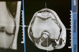
Comparative Techniques in Medical Imaging
In addition to ultrasound, other imaging techniques such as MRI and CT scans can be used to provide more detailed, complementary images. These methods can emphasize tissues that are difficult to distinguish on standard ultrasound due to their isoechoic nature.
Challenges in Identifying Isoechoic Tissue
One of the primary challenges with isoechoic tissues is their potential to mimic normal anatomy. Without proper recognition and additional imaging, such tissues may go undetected. This is particularly vital in cancer detection and monitoring where early identification of masses can significantly impact treatment outcomes.
Advancements in Imaging Technology
Ongoing advancements in imaging technologies aim to tackle the challenges presented by isoechoic structures. Enhanced software algorithms and portable ultrasound devices are among the innovations that help improve diagnostic accuracy.
Training and Experience
The identification of isoechoic tissues often hinges on the technician’s training and experience. Regular training and updating skills with the latest technological advances are crucial for accurate diagnosis. Hands-on workshops and continued education in ultrasound imaging are beneficial in honing these skills.
For more insights on safe and effective medical practices, explore our article on hidden causes of weight issues and effective approaches.
The Future of Diagnosing Isoechoic Structures
The future promises further innovations that will facilitate easier identification of isoechoic structures. AI and machine learning are expected to play a significant role in enhancing imaging accuracy, currently a promising field of research.
Understanding isoechoic structures in medical imaging continues to be a critical aspect of improving diagnostic precision. As technology and methods progress, so does our capability to accurately identify and treat medical conditions. For general health information, consider visiting the comprehensive resources available at Wikipedia.
- Isoechoic structures mimic the echo pattern of surrounding tissues.
- Ultrasound is a primary tool but requires skilled interpretation.
- Complementary imaging techniques enhance detection accuracy.
- Continuous advancements in technology aid in overcoming diagnostic challenges.
- AI and machine learning hold potential for future developments.
What does isoechoic mean in medical imaging?
Isoechoic refers to structures that produce echoes of the same intensity as surrounding tissues in ultrasound imaging, making them challenging to differentiate.
How do isoechoic structures affect diagnosis?
These structures can blend with normal tissues, potentially leading to missed diagnoses if not carefully examined by experienced professionals.
Which imaging techniques can help visualize isoechoic tissues?
Besides ultrasound, MRI and CT scans can provide additional details to distinguish these structures more clearly.
Why is identifying isoechoic structures important?
Recognizing these structures is crucial for accurate diagnosis and successful treatment planning, especially in detecting malignancies or other medical conditions.
Are there advancements that aid in identifying isoechoic structures?
Yes, there are ongoing advancements in imaging technology and techniques that improve the ability to identify these structures accurately, including AI innovations.
Other Articles You May Find of Interest...
- Is an MRI Lumbar Spine Without Contrast Right for You?
- Exploring the Insights of a Normal Shoulder X-Ray
- Is the Lexiscan Stress Test Right for You in Assessing Heart Health?
- Interpreting the Normal Ankle X-Ray: What It Reveals About Your Joint Health
- Exploring the Role of Isoechoic Structures in Medical Imaging
- What Does an Abnormal Chest X-Ray Indicate About Your Health?
- Exploring Hypoattenuation: What It Means for Your Health


