


More Imaging Articles
Detecting Subdural Haematomas: The Role of CT Scans in Diagnosis
Detecting a subdural haematoma can be a critical and time-sensitive process. The role of the subdural haematoma CT scan is indispensable in determining the presence and extent of the condition. CT scans, or computed tomography scans, are a type of imaging that provides detailed pictures of the brain and are often the first step in diagnosing a subdural haematoma.
The Role of Subdural Haematoma CT Scan
A subdural haematoma is a type of bleeding that occurs between the brain and its outer covering, the dura mater. It often results from a head injury, where the blood vessels rupture and blood accumulates, leading to increased pressure on the brain. Identifying this condition quickly is crucial, and CT scans are usually the preferred imaging technique due to their accuracy and speed.
CT scans work by taking numerous X-ray measurements from different angles to reconstruct a comprehensive image of the brain. For suspected subdural haematomas, they reveal whether there is bleeding and help pinpoint its location, size, and potential impact on brain structures. This information is vital in deciding subsequent medical or surgical interventions.
How Are CT Scans Conducted?
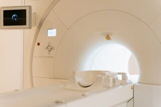
During a CT scan, the patient lies on a motorized table that slides into a cylindrical machine. The process is quick, often taking less than 10 minutes. The scan itself is painless, although patients are required to remain still to ensure clear images. In some cases, a contrast dye is injected to accentuate the images.
Healthcare professionals analyze CT scan results to identify abnormalities in brain structure and any signs of bleeding. Rapid diagnosis through a subdural haematoma CT scan can significantly influence patient outcomes by allowing timely interventions. To understand how early detection of critical conditions impacts health, explore the importance of recognizing early symptoms.
Symptoms Requiring Immediate Attention
Symptoms that may necessitate a CT scan for subdural haematoma include severe headaches, confusion, vomiting, and weakness, usually on one side of the body. These signs, especially after a head injury, should prompt immediate medical evaluation to rule out serious conditions such as a subdural haematoma. Acting swiftly can prevent long-term damage or even be life-saving.
Older adults and individuals on anticoagulant therapy are particularly susceptible to subdural haematomas, even from minor head trauma. Awareness and timely management of symptoms are essential to minimize complications.
Challenges and Considerations
While CT scans are invaluable, they are not without limitations. One of the primary concerns is exposure to radiation, which, although minimal, is an essential consideration, especially for repeated scans. Clinicians weigh the benefits and risks of CT scanning on a case-by-case basis.
Moreover, dense injuries may obscure smaller details on CT images. In such cases, further imaging techniques like MRI may be warranted for comprehensive evaluation. It is crucial to discuss individual circumstances and risks with healthcare providers to make informed decisions.
Advanced Imaging Techniques
In some scenarios, especially when initial imaging results are inconclusive, advanced imaging techniques such as Magnetic Resonance Imaging (MRI) are employed. MRIs use magnetic fields instead of X-rays and can offer more detailed images of brain structures, sometimes better clarifying complex cases that CT scans alone cannot diagnose.
Advanced techniques complement CT scans by providing high-resolution images that might reveal subtle changes. Such integrated approaches can inform a thorough treatment plan.
Prognosis and Treatment
The prognosis for a subdural haematoma depends on the size and impact of the bleed, as well as the timeliness of intervention. Treatment may involve surgical procedures like a craniotomy or burr hole drainage to alleviate pressure on the brain.
Post-surgical rehabilitation might be necessary to address neurological deficits. The aim is to restore function and improve the quality of life. Patients should have regular follow-ups to monitor progress and address any ongoing concerns.
Effective treatment often requires a multidisciplinary team, including neurologists, neurosurgeons, and rehabilitation specialists, ensuring comprehensive care for the patient.
Concluding Thoughts
Understanding and effectively utilizing a subdural haematoma CT scan is critical in diagnosing and managing this potentially life-threatening condition. These scans provide rapid and reliable results, guiding clinical decisions in urgent scenarios. For those wanting more detailed information on cerebral imaging practices, you might find this resource valuable: Medical Imaging on Wikipedia.
A hughlight for the importance of utilizing subdural haematoma CT scans lies in their capacity to affect clinical outcomes positively. Quick and precise imaging enables prompt intervention, significantly impacting recovery chances.
- CT scans are crucial for diagnosing subdural haematomas quickly.
- Subdural haematomas can be life-threatening if not treated promptly.
- Advanced imaging techniques may be necessary for complex cases.
- Immediate medical attention is essential when symptoms of head injury occur.
- Discuss risks and benefits of CT scans with healthcare providers.
FAQ
What is a subdural haematoma?
A subdural haematoma is bleeding that occurs between the brain and its outer covering, often due to head trauma.
Why is a CT scan used to diagnose a subdural haematoma?
CT scans provide fast, detailed cross-sectional images of the brain, allowing healthcare providers to detect bleeding and assess its severity.
Can a subdural haematoma heal on its own?
Small haematomas may resolve independently, but larger ones often require surgical intervention to prevent severe complications.
What symptoms should prompt an immediate CT scan?
Severe headache, confusion, vomiting, and unilateral weakness following head injury warrant immediate medical evaluation, often including a CT scan.
Is a CT scan safe for everyone?
While CT scans involve radiation exposure, they are generally safe. Discuss potential risks with your healthcare provider, especially if repeated scans are necessary.
Other Articles You May Find of Interest...
- MRI vs X-Ray: Which Imaging Technique is Right for You?
- Biparietal Diameter Explained: Understanding Its Importance in Prenatal Care
- What Your Hip X-Ray Reveals About Your Joint Health
- Exploring the Benefits of CT Abdomen and Pelvis With Contrast for Accurate Diagnosis
- Navigating the Insights of MRI for Pituitary Health
- Navigating the Implications of Birads 3 for Your Breast Health
- What Does Hyperechoic Mean and How Does It Impact Your Health?


