More Dental Health Articles
Laser and CT Tech In Your Dental Office
Laser: Highly advanced dental technologies bring the most comfortable and excellent dental experience possible. Most patients do not need to be numb when in-office lasers are used for fillings, and there is no drilling involved depending on the tooth condition. The laser beam removes the decay without removing additional enamel as opposed to the conventional drill, which does take away some tooth structure. For deep cavities that are close to the nerve and run the risk of possibly needing a root canal, the laser reduces that chance by 90%, by eliminating bacteria and desensitizing the tooth. The laser enables dentists to treat your tooth while being very conservative. When the laser is used with a crown prep, it sterilizes the tooth and reduces the chance for root canals by 90%
Nitelase: Used to stimulate collagen and tighten tissues in the back of throat to help open airway to eliminate snoring. Usually patients will need treatment every 21 days for a total of 4-5 treatments and then may need a touch up once per year.
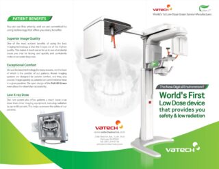
CBCT: What is Green-CT?
- Low X-ray radiation: providing safer CT scans with the latest advances in X-ray technology
- FDA cleared for pediatric use: Child sized X-ray doses, minimizing danger to our children.
- Eco-friendly device: Safer materials and less impacts on environment
The CT image can be used to identify a broad range of malformation, especially around the mouth, jaw, and neck. It’s ability to see fine anatomical structures (in 3D), has become valuable in treating conditions in many areas of dentistry.
- Endodontics: The 3D image can reveal the exact shape and branches of the root canals, as well as the location of the infection. This then allows the dentist to remove the inner infected pulp tissues and replace it with a sterile material.
- Oral Surgery: The 3D image can provide critical information when it comes to impacted tooth and its relationship to adjacent teeth and nerve canals. With this information, proper treatment can then be recommended.
- Dental Implants: For a dental implant to be placed, the tissues of the target area cannot be infected and there needs to be enough bone. The 3D image reveals those factors as well as identifies proper location for implant placement. This increases the success rate of implants.
- Orthodontics: The 3D image can provide information as to where and how the teeth can be moved, especially during growth and development.
- Orthognathic Jaw Surgery: Complex cases that involve both orthodontic tooth movements and orthognathic jaw surgery can be evaluated by using the CT image.
- Temporo-Mandibular Joint (TMJ) Disease: The 3D image can reveal facial pain by viewing the TMJ, teeth, sinuses, and airway.
- Sleep Apnea: The 3D image can reveal the width of the airway, nose, mouth, and windpipe, thus allowing for proper treatment for the patient.
Other Articles You May Find of Interest...
- Let’s Smile Dental’s 7&Up Club
- Strengthening Smiles: Understanding the Importance of Splinting Periodontally Involved Teeth
- Understanding Soft Tissue Grafting: A Key To Periodontal Health
- New Solutions for Dentures and Dental Implants
- Cerec Dental Technology
- Commonly Treated Orthodontic Problems
- Can You Benefit From Braces?

