More Dental Health Articles
Dental Technology: Cone Beam CT Imaging

Cone Beam Computed Tomography (CBCT) promises to change the way many dental problems are diagnosed and treated.
For the dentist, it offers the ability to visualize intricate structures inside the mouth, such as root canals, nerves and sinuses in the jaw – in three dimensions – without surgery.
For the patient, it can reduce the need for invasive procedures, shorten treatment time and offer the chance for a better outcome.
How Cone Beam CT Works
X-rays can penetrate bone and soft tissue, and reveal its hidden structure by their varying degrees of absorption; in other words, they form a grayscale picture of what’s underneath the surface. But conventional X-rays are limited: Like a still-life picture, they show only one perspective on the scene.
Now imagine a “flip book” – the kind of small book made up of a series of pictures, each slightly different. When you rapidly page through it, you may see (for example) an animated cartoon. If you could put together a flip book made from a series of X-ray “slices” of the same subject, taken at slightly different angles, you would be able to create an “animation” of the X-rays. And from there, it’s only one more step to making a 3-D model.
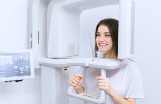
That’s exactly what CBCT scanners do. Using a rotating imaging device that moves around the patient’s head, the scanner records between 150 and 600 different X-ray views in under a minute. Then, a powerful computer processes the information and creates a virtual model of the area under study. When it’s done, the model appears as a three-dimensional image on a computer screen: It can be rotated from side to side or up and down, examined in greater or less detail, and manipulated in any number of ways – all without the patient feeling any discomfort…or even being present.
Where Cone Beam CT Is Used
The ability to see fine anatomical structures in 3-D has proven invaluable in treating conditions in many areas of dentistry.
- Orthodontics: Helps determine exactly how and where teeth should be moved.
- Dental Implants: Used to determine the optimum location for the titanium implants while avoiding nerves, sinuses and areas of low bone density.
- Orthognathic Jaw Surgery and TMJ: Patients benefit when the specialists who treat these conditions can evaluate their anatomy with the three-dimensional perspective that cone beam CT provides.
- Oral Surgery: Treatment for tumors or impacted teeth is aided by the level of fine detail shown in these scans.
- Endodontics: Dentists performing intricate procedures (like complex root canals) benefit from a clearer visualization of the tooth’s anatomy.
- Sleep Apnea: Imaging the tissues and structures of the nose, mouth and throat can aid in diagnosis and treatment of this dangerous condition.
CBCT changes the way many dental problems are diagnosed and treated. To find out how it can benefit you schedule an appointment today!
Other Articles You May Find of Interest...
- Let’s Smile Dental’s 7&Up Club
- Strengthening Smiles: Understanding the Importance of Splinting Periodontally Involved Teeth
- Understanding Soft Tissue Grafting: A Key To Periodontal Health
- New Solutions for Dentures and Dental Implants
- Benefits Of Immediate Dental Implants
- Preventing Tooth Injuries During Your Child’s Active Summer
- How New Tech In the Dental Office Benefits You


