


More Health Technology Articles
The Evolution of Modern Microscopy in Scientific Research
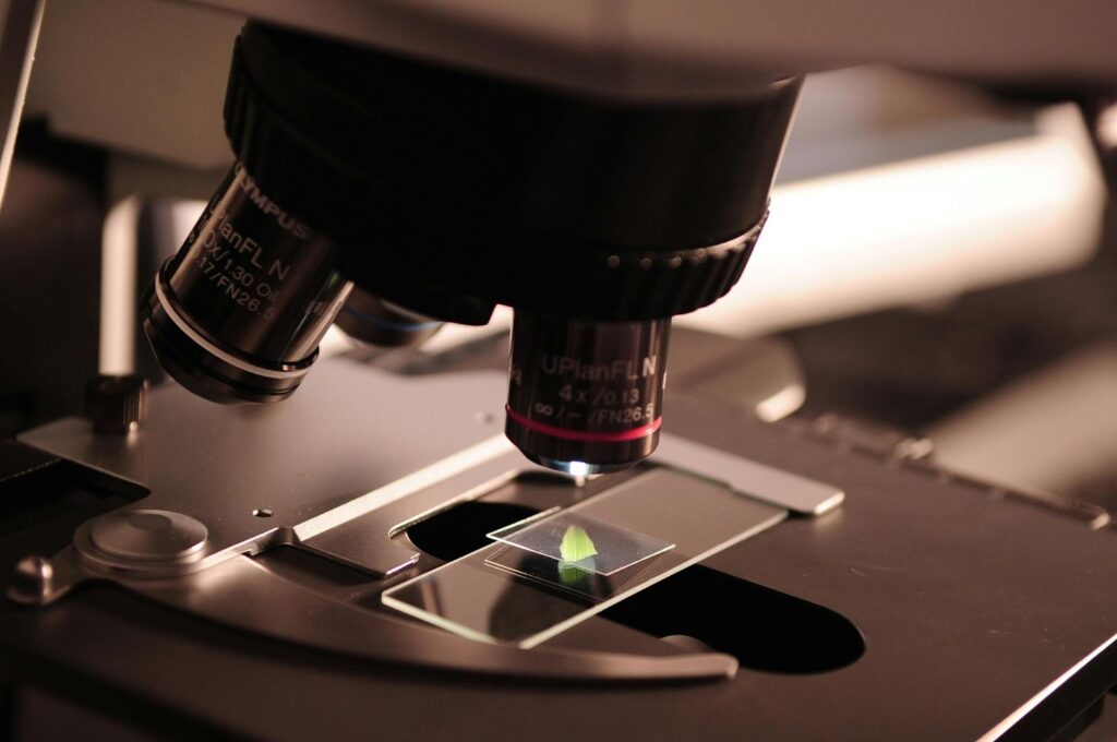
Microscopes have totally changed how scientists peek into the world. These gadgets let researchers zoom in on things we can’t see with just our eyes. Modern microscopy’s journey has sparked some jaw-dropping leaps in biology, medicine, and materials science. From clunky old-school lenses to today’s high-tech imaging rigs, it’s a field that just keeps growing.
Early Developments in Microscopy
Way back in the late 16th century, the first microscopes popped up. They were pretty basic—just simple lenses that made things a bit bigger, though not super clear. Then came Antonie van Leeuwenhoek, this Dutch guy who tweaked the design and spotted single-celled critters for the first time. Talk about a game-changer—he basically kicked off microbiology. Those early scopes weren’t fancy, but they cracked open a whole new world for curious minds.
Over the 17th and 18th centuries, folks got smarter with optical lenses, bumping up magnification and sharpness. Compound microscopes, with their multi-lens setup, started showing up more. Scientists could now check out bacteria, plant cells, even tiny bugs in way more detail. Still, these tools had their limits—fuzzy resolution and weak light sources held things back. Innovations in glass manufacturing gradually helped fix the blurry edges that often plagued early scientific observations. Makers started crafting high-quality microscope objectives that could gather more light and sharpen the tiny details of a sample. This shift toward precision optics paved the way for the massive breakthroughs seen in modern research facilities.
The Rise of Fluorescence Microscopes
By the 20th century, researchers were itching for something more reliant to study tiny structures. Enter fluorescence microscopy—a total breakthrough. Fluorescence microscopes use fluorescent dyes to highlight specific structures. Shine the right light on them, and they’d glow, spotlighting whatever you wanted to see.
That shift flipped biological research on its head. Scientists could zero in on proteins, DNA, and cell parts with crisp detail. It became a go-to for medical diagnostics too, sniffing out diseases at the molecular level. Immunofluorescence let folks watch cells in action, live. Even now, it’s a heavyweight in biomedical labs.
Electron Microscopy and High-Resolution Imaging
Optical microscopes got better, sure, but they hit a wall—light’s wavelength just couldn’t cut it for the tiniest things. Then electron microscopy swooped in, swapping light for electron beams. The result? Insane magnification and resolution that left old-school scopes in the dust.
These beasts let researchers eyeball viruses, organelles, and molecular details like never before. Two big players came out of it: transmission electron microscopy (TEM) for crazy-detailed insides, and scanning electron microscopy (SEM) for cool 3D surface shots. Biology got a boost with close-ups of cell membranes, while materials science dove into metals and nanomaterials. Electron scopes keep pushing the envelope across all kinds of fields.
The Emergence of Confocal Microscopy
Old optical microscopes grabbed light from everywhere at once—great, until your image turned into a blurry mess, especially with chunky samples. Confocal microscopy fixed that by zapping a laser at one depth at a time. It kicked stray light to the curb and sharpened things up.
That trick let scientists build 3D cell and tissue pics. Neuroscientists loved it for mapping brain bits, and cancer researchers tracked tumor moves with it. By slicing through depths with crisp focus, confocal scopes made living cell studies way clearer—no more fuzzy nonsense. It’s still a rock star in bio research.
Super-Resolution Microscopy and Breaking Limits
For ages, folks thought light microscopes had hit their peak resolution—done deal. Then super-resolution microscopy crashed the party. These techniques blew past old limits, letting researchers peek at molecular-scale action.
Stimulated Emission Depletion (STED) used laser wizardry to tighten fluorescence focus, while Photoactivated Localization Microscopy (PALM) played with molecule glows for ultra-sharp shots. Cell biology went nuts—tracking proteins and interactions got real precise. It’s shed light on Alzheimer’s, cancer, even infections. Seeing the nanoscale like this keeps rewriting what we know.
The Role of AI and Digital Microscopy
Now artificial intelligence is shaking things up. Digital microscopes with AI smarts can chew through images on their own. That speed’s a godsend—spotting patterns or weird occurrences in a snap. It’s a whiz at diagnosing diseases, testing drugs, even cleaning up fuzzy pics.
Machine learning’s cutting noise and boosting clarity too. In medicine, it’s catching cancer cells with eagle-eye accuracy. Materials science leans on it to flag nanomaterial flaws. Mixing AI with digital imaging? That’s a frontier-pushing combo right there.

Future Trends in Microscopy
Microscopy’s future looks wild. Real-time imaging’s on the horizon—think instant breakdowns of bio and chemical action. Quantum microscopy’s bubbling up too, promising sharper, more sensitive views. Toss in nanotech and optical upgrades, and we’re headed for some next-level gear.
From those first rickety lenses to AI-powered setups, microscopy’s come a long haul. Every jump forward stretches what we can see. It’s been a powerhouse for discovery in medicine, biology, and materials—and it’s not slowing down anytime soon.
Other Articles You May Find of Interest...
- Healthcare’s Next Efficiency Revolution: Wearable Apps, Real-Time Data, and EHR Integration
- Smart Health Habits: Using Technology to Support a Healthier Lifestyle
- The Future of Patient Care and the Technology Making It Possible
- The AI-Enabled Entrepreneur: How Technology Is Lowering Barriers to Starting a Business
- AI Wellness Apps vs Traditional Meditation Apps in 2026
- Building Resilient Healthcare Systems Through Proactive Cybersecurity Strategies
- Healthcare Call Centers Improving Patient Communication in 2026


