More Beauty Skin Care & Gorgeous Smiles Articles
What Happens Behind a Skin Biopsy
A skin biopsy feels routine from the patient’s side. A small procedure, a bandage, instructions to wait for results. What happens between the moment that tissue sample leaves the clinic and the moment a pathology report reaches a physician is a process most patients never think about — and one that determines whether everything that follows is built on solid ground.
The work is meticulous, multi-step, and depends on both technical precision and interpretive judgment. Understanding it doesn’t change the patient experience. It does explain why the waiting period exists, and why accuracy in this process matters in ways that cutting corners simply can’t accommodate.
From Clinic to Laboratory
The journey starts at collection. How a biopsy sample gets handled in the minutes after removal affects the quality of what the laboratory receives — sometimes significantly. Proper fixation in formalin preserves the cellular structure pathologists need to evaluate. Samples that are poorly fixed, mislabeled, or delayed in transit can compromise the analysis before any laboratory work has even begun.
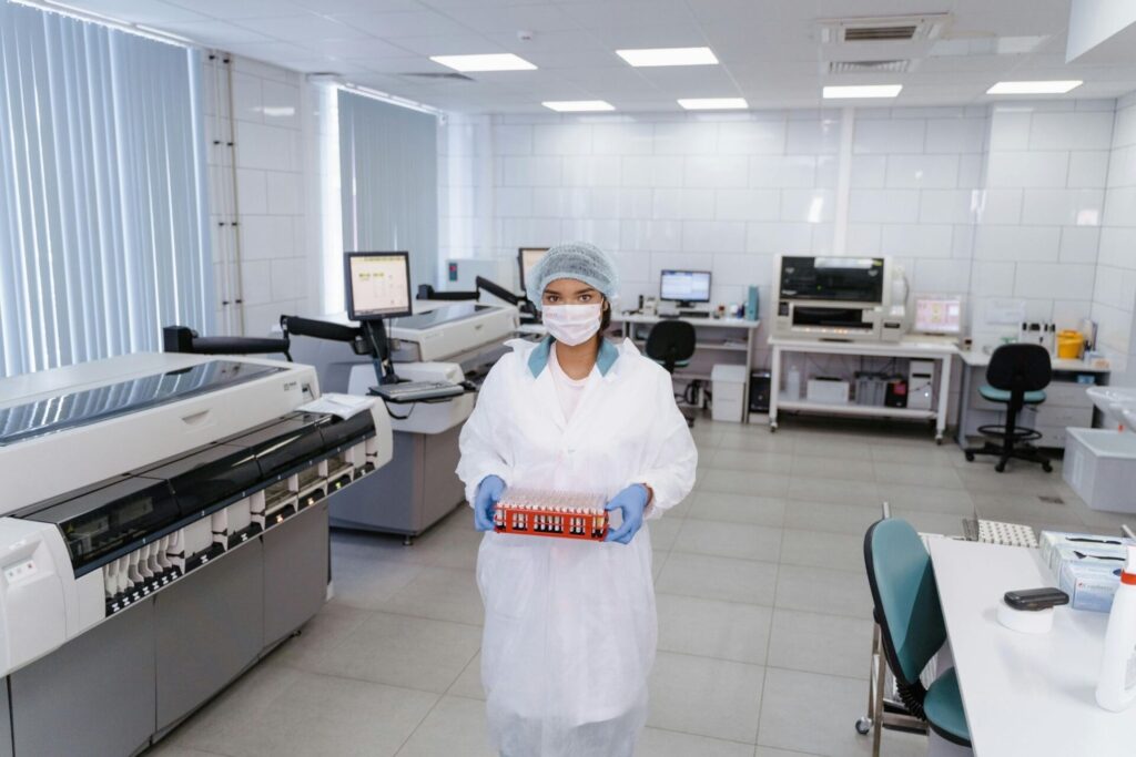
Once it arrives, the sample gets logged, assigned a case number, and enters a processing workflow that will take it from a small piece of tissue to a set of glass slides ready for microscopic examination. That workflow involves considerably more steps than most people would guess.
Tissue Processing and Embedding
Before the tissue can be sectioned, it has to be dehydrated, cleared of fixative, and infiltrated with paraffin wax. This processing cycle typically runs overnight. After that comes embedding — a technician orients the tissue precisely in a paraffin block, and that orientation matters more than it sounds. The plane the tissue sits in determines what the pathologist will actually be able to see when the sections are cut.
This step requires judgment, not just technique. A technician who understands how different tissue types behave, and who recognizes when something needs special handling, affects the quality of the final result in ways that never appear in the report itself. It’s invisible work with visible consequences.
Sectioning and Staining
The paraffin block goes onto a microtome — an instrument that cuts sections at around four micrometers thick, a fraction of the width of a human hair. Those sections get floated onto glass slides, dried, and stained. The standard stain is hematoxylin and eosin, which gives cell nuclei a blue-purple color and cytoplasm a pink one. That color contrast is what allows pathologists to distinguish structures that would otherwise be invisible.
Some cases need more than H&E. Immunohistochemical stains use antibodies to detect specific proteins in the tissue — useful for classifying tumors, identifying infectious organisms, or resolving diagnostic questions that routine staining leaves open. Not every case needs them, and deciding which cases do requires interpretive judgment before the pathologist ever looks at the slides.
That’s where the case-specific thinking starts to matter. PathScience, for example, applies ancillary testing based on what an individual case actually warrants rather than defaulting to a standard panel regardless of clinical context — a distinction that affects both efficiency and diagnostic accuracy in ways that compound across a high volume of cases.
The Pathologist’s Review
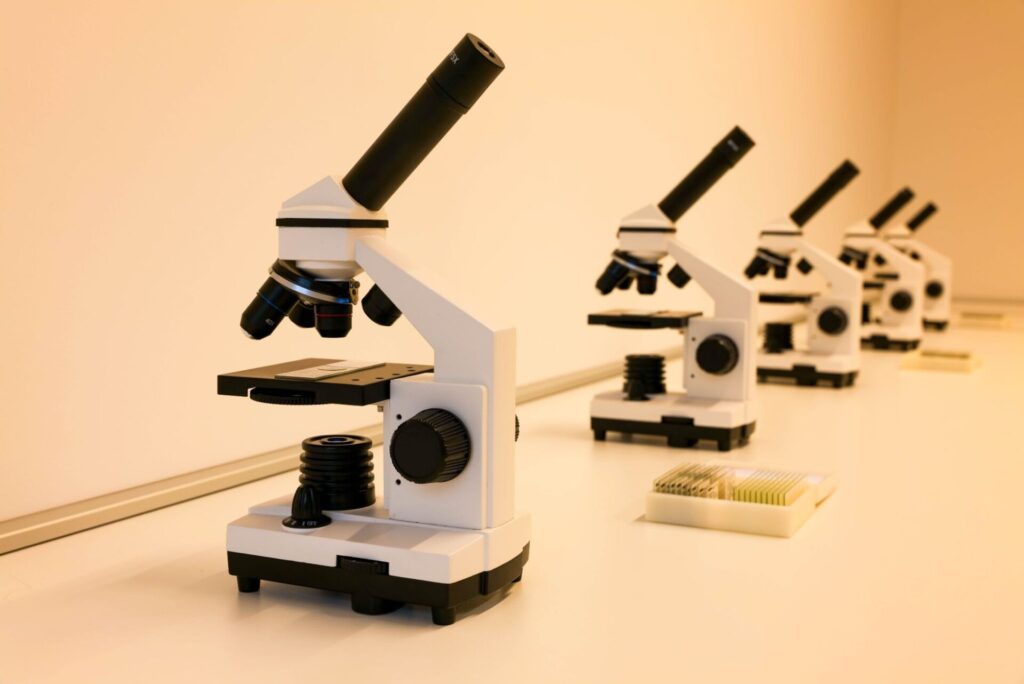
With slides prepared, the case reaches the pathologist. What happens at the microscope goes well beyond visual inspection. The pathologist is integrating what they see in the tissue with the clinical information the submitting physician provided — patient age, lesion location, how long it’s been present, what it looked like at the time of biopsy.
Context shapes interpretation. The same histological finding can mean something different depending on the clinical picture, and experienced pathologists weigh that actively. It’s not a passive process.
Straightforward cases move relatively quickly. Cases with unusual features, overlapping patterns, or findings that don’t fit a single category cleanly take longer — additional stains, a colleague consultation, a second pass through the literature. The time that takes is part of why results aren’t always immediate.
The Report
The pathology report is where everything gets translated into language a treating physician can act on. A useful report doesn’t just state a diagnosis. It provides the specific information needed to make decisions about treatment, staging, and follow-up.
For excised lesions, margin status matters. For melanocytic lesions, tumor thickness and mitotic rate inform surgical planning and oncological management. The level of detail in a report reflects the level of analysis behind it — and clinicians who have worked with both thorough and superficial reports know the difference in how confidently they can move forward.
Why Any of This Matters to the Patient
The patient waiting on a biopsy result is at the beginning of a clinical decision tree. What gets recommended next — treatment, surveillance, reassurance, urgency — traces directly back to what the pathology report says and how specifically it says it.
None of the process described above is visible from the waiting room. But it’s present in every result that comes back accurate, specific, and useful to the physician who has to decide what to do next. That’s what the work is for.
Other Articles You May Find of Interest...
- Botox vs Dysport vs Xeomin: Which Neuromodulator Is Right for You?
- Understanding PDRN Therapy: The Science Behind Skin Barrier Repair and Tissue Regeneration
- Who Is a Suitable Candidate for Breast Augmentation?
- Peptides vs Retinoids for Sensitive Skin: Why Matrixyl 3000 and CoQ10 Are Changing the Conversation
- Whitehead or Pustule? How to Tell These Acne Bumps Apart
- What Patients Should Know About Skin Removal Surgery
- Breast Augmentation vs. Breast Lift: Understanding the Difference